 Movie
Movie Controller

[English] 日本語
 Yorodumi
- PDB-3mf7: Crystal Structure of (R)-oxirane-2-carboxylate inhibited cis-CaaD -
+ Open data

- Basic information
| Entry | Database: PDB / ID: 3mf7 | ||||||
|---|---|---|---|---|---|---|---|
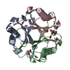
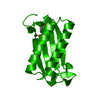
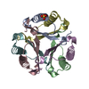
| Title | Crystal Structure of (R)-oxirane-2-carboxylate inhibited cis-CaaD | ||||||
 Components Components | Cis-3-chloroacrylic acid dehalogenase | ||||||
 Keywords Keywords | HYDROLASE / beta-alpha-beta motif / tautomerase / dehalogenase / cis-3-chloroacrylic acid dehalogenase / Isomerase | ||||||
| Function / homology | Tautomerase, cis-CaaD-like / Putative oxalocrotonate tautomerase enzyme / Macrophage Migration Inhibitory Factor / Macrophage Migration Inhibitory Factor / Tautomerase/MIF superfamily / 2-Layer Sandwich / Alpha Beta / Cis-3-chloroacrylic acid dehalogenase Function and homology information Function and homology information | ||||||
| Biological species |  coryneform bacterium (bacteria) coryneform bacterium (bacteria) | ||||||
| Method |  X-RAY DIFFRACTION / MOLECULAR REPLACEMENT / Resolution: 1.65 Å X-RAY DIFFRACTION / MOLECULAR REPLACEMENT / Resolution: 1.65 Å | ||||||
 Authors Authors | Guo, Y. / Serrano, H. / Ernst, S.R. / Johnson Jr., W.H. / Hackert, M.L. / Whitman, C.P. | ||||||
 Citation Citation | Journal: Bioorg.Chem. / Year: 2011 Title: Crystal structures of native and inactivated cis-3-chloroacrylic acid dehalogenase: Implications for the catalytic and inactivation mechanisms. Authors: Guo, Y. / Serrano, H. / Johnson, W.H. / Ernst, S. / Hackert, M.L. / Whitman, C.P. | ||||||
| History |
|
- Structure visualization
| Structure viewer | Molecule: MolmilJmol/JSmol |
|---|
- Downloads & links
-Download
| PDBx/mmCIF format | 3mf7.cif.gz | 62.9 KB | Display | PDBx/mmCIF format |
|---|---|---|---|---|
| PDB format | pdb3mf7.ent.gz | 45.7 KB | Display | PDB format |
| PDBx/mmJSON format | 3mf7.json.gz | Tree view | PDBx/mmJSON format | |
| Others |  Other downloads Other downloads |
-Validation report
| Summary document | 3mf7_validation.pdf.gz | 430.7 KB | Display | wwPDB validaton report |
|---|---|---|---|---|
| Full document | 3mf7_full_validation.pdf.gz | 431.7 KB | Display | |
| Data in XML | 3mf7_validation.xml.gz | 8.7 KB | Display | |
| Data in CIF | 3mf7_validation.cif.gz | 11.4 KB | Display | |
| Arichive directory | https://data.pdbj.org/pub/pdb/validation_reports/mf/3mf7ftp://data.pdbj.org/pub/pdb/validation_reports/mf/3mf7 | HTTPS FTP |
-Related structure data
| Related structure data | 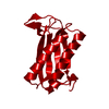 3mf8SC S: Starting model for refinement C: citing same article ( |
|---|---|
| Similar structure data |


-Links
 PDBj
PDBj
- Assembly
| Deposited unit | 
| |||||||||
|---|---|---|---|---|---|---|---|---|---|---|
| 1 | 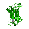
| |||||||||
| Unit cell |
| |||||||||
| Components on special symmetry positions |
|
-Components
| #1: Protein | Mass: 16732.590 Da / Num. of mol.: 1 / Fragment: UNP residues 2-118 Source method: isolated from a genetically manipulated source Source: (gene. exp.) coryneform bacterium (bacteria) / Gene: cis-caaD / Plasmid: pET3 / Production host: References: UniProt: Q6VPE5, Hydrolases; Acting on halide bonds; In carbon-halide compounds |
|---|---|
| #2: Water | ChemComp-HOH /  Mass: 18.015 Da / Num. of mol.: 117 / Source method: isolated from a natural source / Formula: H2O Mass: 18.015 Da / Num. of mol.: 117 / Source method: isolated from a natural source / Formula: H2O |
-Experimental details
-Experiment
| Experiment | Method: X-RAY DIFFRACTION / Number of used crystals: 1 |
|---|
- Sample preparation
| Crystal | Density Matthews: 2.26 Å3/Da / Density % sol: 45.51 % |
|---|---|
| Crystal grow | Temperature: 298 K / Method: hanging drop / pH: 4.6 Details: three micro liter of 15.6mg/mL cis-CaaD protein sample mixed with three micro liter crystallization solution: 0.125 M CaCl2, 0.07 M sodium acetate buffer, 12.5% isopropanol, hanging drop, ...Details: three micro liter of 15.6mg/mL cis-CaaD protein sample mixed with three micro liter crystallization solution: 0.125 M CaCl2, 0.07 M sodium acetate buffer, 12.5% isopropanol, hanging drop, temperature 298K, pH 4.6 |
-Data collection
| Diffraction | Mean temperature: 100 K | ||||||||||||||||||||||||||||||||||||||||||||||||||||||||||||||||||
|---|---|---|---|---|---|---|---|---|---|---|---|---|---|---|---|---|---|---|---|---|---|---|---|---|---|---|---|---|---|---|---|---|---|---|---|---|---|---|---|---|---|---|---|---|---|---|---|---|---|---|---|---|---|---|---|---|---|---|---|---|---|---|---|---|---|---|---|
| Diffraction source | Source: ROTATING ANODE / Type: RIGAKU RU200 / Wavelength: 1.5418 Å | ||||||||||||||||||||||||||||||||||||||||||||||||||||||||||||||||||
| Detector | Type: RIGAKU RAXIS / Detector: IMAGE PLATE / Date: Apr 25, 2005 | ||||||||||||||||||||||||||||||||||||||||||||||||||||||||||||||||||
| Radiation | Monochromator: BLUE MAX-FLUX OPTICAL SYSTEM / Protocol: SINGLE WAVELENGTH / Monochromatic (M) / Laue (L): M / Scattering type: x-ray | ||||||||||||||||||||||||||||||||||||||||||||||||||||||||||||||||||
| Radiation wavelength | Wavelength: 1.5418 Å / Relative weight: 1 | ||||||||||||||||||||||||||||||||||||||||||||||||||||||||||||||||||
| Reflection | Resolution: 1.62→50 Å / Num. obs: 19123 / % possible obs: 98.9 % / Redundancy: 4.6 % / Rmerge(I) obs: 0.063 / Χ2: 3.096 / Net I/σ(I): 24 | ||||||||||||||||||||||||||||||||||||||||||||||||||||||||||||||||||
| Reflection shell |
|
- Processing
| Software |
| ||||||||||||||||||||||||||||||||||||||||||||||||||||||||||||||||||||||||||||||||||||||||||
|---|---|---|---|---|---|---|---|---|---|---|---|---|---|---|---|---|---|---|---|---|---|---|---|---|---|---|---|---|---|---|---|---|---|---|---|---|---|---|---|---|---|---|---|---|---|---|---|---|---|---|---|---|---|---|---|---|---|---|---|---|---|---|---|---|---|---|---|---|---|---|---|---|---|---|---|---|---|---|---|---|---|---|---|---|---|---|---|---|---|---|---|
| Refinement | Method to determine structure: MOLECULAR REPLACEMENT Starting model: PDB entry 3MF8 Resolution: 1.65→50 Å / Cor.coef. Fo:Fc: 0.939 / Cor.coef. Fo:Fc free: 0.911 / Occupancy max: 1 / Occupancy min: 0 / SU B: 3.282 / SU ML: 0.052 / Cross valid method: THROUGHOUT / σ(F): 0 / ESU R: 0.115 / ESU R Free: 0.093 / Stereochemistry target values: MAXIMUM LIKELIHOOD / Details: HYDROGENS HAVE BEEN ADDED IN THE RIDING POSITIONS
| ||||||||||||||||||||||||||||||||||||||||||||||||||||||||||||||||||||||||||||||||||||||||||
| Solvent computation | Ion probe radii: 0.8 Å / Shrinkage radii: 0.8 Å / VDW probe radii: 1.4 Å / Solvent model: MASK | ||||||||||||||||||||||||||||||||||||||||||||||||||||||||||||||||||||||||||||||||||||||||||
| Displacement parameters | Biso max: 41.24 Å2 / Biso mean: 9.866 Å2 / Biso min: 2 Å2 | ||||||||||||||||||||||||||||||||||||||||||||||||||||||||||||||||||||||||||||||||||||||||||
| Refinement step | Cycle: LAST / Resolution: 1.65→50 Å
| ||||||||||||||||||||||||||||||||||||||||||||||||||||||||||||||||||||||||||||||||||||||||||
| Refine LS restraints |
| ||||||||||||||||||||||||||||||||||||||||||||||||||||||||||||||||||||||||||||||||||||||||||
| LS refinement shell | Resolution: 1.65→1.693 Å / Total num. of bins used: 20
|
