 Movie
Movie Controller

[English] 日本語
 Yorodumi
- PDB-3mdi: Crystal Structure of the 25kDa Subunit of Human Cleavage factor I... -
+ Open data

- Basic information
| Entry | Database: PDB / ID: 3mdi | ||||||
|---|---|---|---|---|---|---|---|
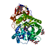
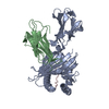
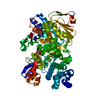
| Title | Crystal Structure of the 25kDa Subunit of Human Cleavage factor Im in complex with RNA UGUAAA | ||||||
 Components Components |
| ||||||
 Keywords Keywords | RNA binding/RNA / CPSF5 / CF Im / mRNA processing / cleavage factor / Nudix protein / protein-RNA complex / RNA binding protein / RNA binding-RNA complex | ||||||
| Function / homology |  Function and homology information Function and homology informationpositive regulation of pro-B cell differentiation / mRNA cleavage factor complex / co-transcriptional mRNA 3'-end processing, cleavage and polyadenylation pathway / Processing of Intronless Pre-mRNAs / mRNA cleavage and polyadenylation specificity factor complex / mRNA alternative polyadenylation / positive regulation of stem cell differentiation / mRNA 3'-UTR AU-rich region binding / mRNA 3'-end processing / mRNA 3'-end processing ...positive regulation of pro-B cell differentiation / mRNA cleavage factor complex / co-transcriptional mRNA 3'-end processing, cleavage and polyadenylation pathway / Processing of Intronless Pre-mRNAs / mRNA cleavage and polyadenylation specificity factor complex / mRNA alternative polyadenylation / positive regulation of stem cell differentiation / mRNA 3'-UTR AU-rich region binding / mRNA 3'-end processing / mRNA 3'-end processing / paraspeckles / RNA Polymerase II Transcription Termination / post-transcriptional regulation of gene expression / protein heterotetramerization / Processing of Capped Intron-Containing Pre-mRNA / protein tetramerization / centriolar satellite / histone deacetylase binding / mRNA processing / cell differentiation / nuclear body / mRNA binding / centrosome / chromatin binding / protein homodimerization activity / RNA binding / nucleoplasm / identical protein binding / nucleus / cytoplasm Similarity search - Function | ||||||
| Biological species |  Homo sapiens (human) Homo sapiens (human) | ||||||
| Method |  X-RAY DIFFRACTION / MOLECULAR REPLACEMENT / Resolution: 2.07 Å X-RAY DIFFRACTION / MOLECULAR REPLACEMENT / Resolution: 2.07 Å | ||||||
 Authors Authors | Yang, Q. / Gilmartin, G.M. / Doublie, S. | ||||||
 Citation Citation | Journal: Proc.Natl.Acad.Sci.USA / Year: 2010 Title: Structural basis of UGUA recognition by the Nudix protein CFI(m)25 and implications for a regulatory role in mRNA 3' processing. Authors: Yang, Q. / Gilmartin, G.M. / Doublie, S. | ||||||
| History |
|
- Structure visualization
| Structure viewer | Molecule: MolmilJmol/JSmol |
|---|
- Downloads & links
-Download
| PDBx/mmCIF format | 3mdi.cif.gz | 107.6 KB | Display | PDBx/mmCIF format |
|---|---|---|---|---|
| PDB format | pdb3mdi.ent.gz | 80.1 KB | Display | PDB format |
| PDBx/mmJSON format | 3mdi.json.gz | Tree view | PDBx/mmJSON format | |
| Others |  Other downloads Other downloads |
-Validation report
| Summary document | 3mdi_validation.pdf.gz | 471.2 KB | Display | wwPDB validaton report |
|---|---|---|---|---|
| Full document | 3mdi_full_validation.pdf.gz | 478.2 KB | Display | |
| Data in XML | 3mdi_validation.xml.gz | 20.9 KB | Display | |
| Data in CIF | 3mdi_validation.cif.gz | 29.6 KB | Display | |
| Arichive directory | https://data.pdbj.org/pub/pdb/validation_reports/md/3mdiftp://data.pdbj.org/pub/pdb/validation_reports/md/3mdi | HTTPS FTP |
-Related structure data
| Related structure data |  3mdgC  3bhoS C: citing same article ( S: Starting model for refinement |
|---|---|
| Similar structure data |


-Links
 PDBj
PDBj

- Assembly
| Deposited unit | 
| ||||||||
|---|---|---|---|---|---|---|---|---|---|
| 1 |
| ||||||||
| Unit cell |
|
-Components
| #1: Protein | Mass: 26259.242 Da / Num. of mol.: 2 Source method: isolated from a genetically manipulated source Source: (gene. exp.) Homo sapiens (human) / Gene: CFIM25, CPSF25, CPSF5, NUDT21 / Plasmid: His6-MBP fusion vector / Production host:  #2: RNA chain | | Mass: 1900.197 Da / Num. of mol.: 1 / Source method: obtained synthetically #3: Chemical |   Mass: 92.094 Da / Num. of mol.: 2 / Source method: obtained synthetically / Formula: C3H8O3 Mass: 92.094 Da / Num. of mol.: 2 / Source method: obtained synthetically / Formula: C3H8O3#4: Water | ChemComp-HOH / |  Mass: 18.015 Da / Num. of mol.: 277 / Source method: isolated from a natural source / Formula: H2O Mass: 18.015 Da / Num. of mol.: 277 / Source method: isolated from a natural source / Formula: H2O |
|---|
-Experimental details
-Experiment
| Experiment | Method: X-RAY DIFFRACTION / Number of used crystals: 1 |
|---|
- Sample preparation
| Crystal | Density Matthews: 2.27 Å3/Da / Density % sol: 45.84 % |
|---|---|
| Crystal grow | Temperature: 298 K / Method: vapor diffusion, hanging drop / pH: 4.9 Details: 25% PEG 8000, 0.2M Sodium phosphate pH 4.9, 0.1M Sodium acetate pH 5.0, vapor diffusion, hanging drop, temperature 298K |
-Data collection
| Diffraction | Mean temperature: 100 K |
|---|---|
| Diffraction source | Source: ROTATING ANODE / Type: RIGAKU RU300 / Wavelength: 1.5418 Å |
| Detector | Type: MAR scanner 345 mm plate / Detector: IMAGE PLATE / Date: Jul 31, 2009 / Details: mirrors |
| Radiation | Monochromator: MAR mirrors / Protocol: SINGLE WAVELENGTH / Monochromatic (M) / Laue (L): M / Scattering type: x-ray |
| Radiation wavelength | Wavelength: 1.5418 Å / Relative weight: 1 |
| Reflection twin | Operator: k,h,-l / Fraction: 0.074 |
| Reflection | Resolution: 2.07→20 Å / Num. obs: 30752 / % possible obs: 99.511 % / Redundancy: 6.8 % / Biso Wilson estimate: 31.081 Å2 / Rmerge(I) obs: 0.104 / Net I/σ(I): 14.82 |
| Reflection shell | Resolution: 2.07→2.15 Å / Redundancy: 5.9 % / Rmerge(I) obs: 0.546 / Mean I/σ(I) obs: 3.2 / Num. measured obs: 35077 / Num. unique all: 6307 / Num. unique obs: 6086 / % possible all: 87.3 |
- Processing
| Software |
| |||||||||||||||||||||||||||||||||||||||||||||||||||||||||||||||||||||||||||||||||||||||||||||||||||||||||||||||||||||||||||||||||||||||||||||||||||
|---|---|---|---|---|---|---|---|---|---|---|---|---|---|---|---|---|---|---|---|---|---|---|---|---|---|---|---|---|---|---|---|---|---|---|---|---|---|---|---|---|---|---|---|---|---|---|---|---|---|---|---|---|---|---|---|---|---|---|---|---|---|---|---|---|---|---|---|---|---|---|---|---|---|---|---|---|---|---|---|---|---|---|---|---|---|---|---|---|---|---|---|---|---|---|---|---|---|---|---|---|---|---|---|---|---|---|---|---|---|---|---|---|---|---|---|---|---|---|---|---|---|---|---|---|---|---|---|---|---|---|---|---|---|---|---|---|---|---|---|---|---|---|---|---|---|---|---|---|
| Refinement | Method to determine structure: MOLECULAR REPLACEMENT Starting model: 3BHO Resolution: 2.07→19.937 Å / Occupancy max: 1 / Occupancy min: 0 / FOM work R set: 0.821 / Cross valid method: THROUGHOUT / σ(F): 1.55 / Stereochemistry target values: TWIN_LSQ_F / Details: Twin law: k,h,-l, Twin percentage 0.074
| |||||||||||||||||||||||||||||||||||||||||||||||||||||||||||||||||||||||||||||||||||||||||||||||||||||||||||||||||||||||||||||||||||||||||||||||||||
| Solvent computation | Shrinkage radii: 0.9 Å / VDW probe radii: 1.11 Å / Solvent model: FLAT BULK SOLVENT MODEL / Bsol: 48.575 Å2 / ksol: 0.399 e/Å3 | |||||||||||||||||||||||||||||||||||||||||||||||||||||||||||||||||||||||||||||||||||||||||||||||||||||||||||||||||||||||||||||||||||||||||||||||||||
| Displacement parameters | Biso max: 67.27 Å2 / Biso mean: 26.139 Å2 / Biso min: 10.76 Å2
| |||||||||||||||||||||||||||||||||||||||||||||||||||||||||||||||||||||||||||||||||||||||||||||||||||||||||||||||||||||||||||||||||||||||||||||||||||
| Refinement step | Cycle: LAST / Resolution: 2.07→19.937 Å
| |||||||||||||||||||||||||||||||||||||||||||||||||||||||||||||||||||||||||||||||||||||||||||||||||||||||||||||||||||||||||||||||||||||||||||||||||||
| Refine LS restraints |
| |||||||||||||||||||||||||||||||||||||||||||||||||||||||||||||||||||||||||||||||||||||||||||||||||||||||||||||||||||||||||||||||||||||||||||||||||||
| LS refinement shell | Refine-ID: X-RAY DIFFRACTION / Total num. of bins used: 20
|
