[English] 日本語
Yorodumi
- PDB-3mcr: Crystal structure of NADH dehydrogenase subunit C (Tfu_2693) from... -

+
Open data


ID or keywords:

Loading...

-
Basic information

Entry
Database: PDB / ID: 3mcr
TitleCrystal structure of NADH dehydrogenase subunit C (Tfu_2693) from THERMOBIFIDA FUSCA YX-ER1 at 2.65 A resolution
ComponentsNADH dehydrogenase, subunit C
KeywordsOXIDOREDUCTASE / Structural Genomics / Joint Center for Structural Genomics / JCSG / Protein Structure Initiative / PSI-2
Function / homology
Function and homology information


Translocases; Catalysing the translocation of protons; Linked to oxidoreductase reactions / NADH dehydrogenase (ubiquinone) activity / quinone binding / plasma membrane
Similarity search - Function
NADH:ubiquinone oxidoreductase Nqo5 subunit / NADH dehydrogenase, subunit C / NADH:ubiquinone oxidoreductase, 30kDa subunit / NADH:ubiquinone oxidoreductase, 30kDa subunit superfamily / Respiratory-chain NADH dehydrogenase, 30 Kd subunit / Beta Polymerase; domain 2 / 2-Layer Sandwich / Alpha Beta
Similarity search - Domain/homology
: / HEXANE-1,6-DIOL / NADH-quinone oxidoreductase subunit C
Similarity search - Component
Biological speciesThermobifida fusca (bacteria)
MethodX-RAY DIFFRACTION / SYNCHROTRON / molecular replacement / Resolution: 2.65 Å
AuthorsJoint Center for Structural Genomics (JCSG)
CitationJournal: To be published
Title: Crystal structure of NADH dehydrogenase subunit C (Tfu_2693) from THERMOBIFIDA FUSCA YX-ER1 at 2.65 A resolution
Authors: Joint Center for Structural Genomics (JCSG)
History
DepositionMar 29, 2010Deposition site: RCSB / Processing site: RCSB
Revision 1.0May 12, 2010Provider: repository / Type: Initial release
Revision 1.1Jul 13, 2011Group: Advisory / Version format compliance
Revision 1.2Nov 8, 2017Group: Refinement description / Category: software / Item: _software.classification / _software.name
Revision 1.3Jul 17, 2019Group: Data collection / Refinement description / Category: software
Item: _software.classification / _software.contact_author ..._software.classification / _software.contact_author / _software.contact_author_email / _software.language / _software.location / _software.name / _software.type / _software.version
Revision 1.4Feb 1, 2023Group: Database references / Derived calculations
Category: database_2 / pdbx_struct_conn_angle ...database_2 / pdbx_struct_conn_angle / struct_conn / struct_ref_seq_dif / struct_site
Item: _database_2.pdbx_DOI / _database_2.pdbx_database_accession ..._database_2.pdbx_DOI / _database_2.pdbx_database_accession / _pdbx_struct_conn_angle.ptnr1_auth_comp_id / _pdbx_struct_conn_angle.ptnr1_auth_seq_id / _pdbx_struct_conn_angle.ptnr1_label_atom_id / _pdbx_struct_conn_angle.ptnr1_label_comp_id / _pdbx_struct_conn_angle.ptnr1_label_seq_id / _pdbx_struct_conn_angle.ptnr3_auth_comp_id / _pdbx_struct_conn_angle.ptnr3_auth_seq_id / _pdbx_struct_conn_angle.ptnr3_label_atom_id / _pdbx_struct_conn_angle.ptnr3_label_comp_id / _pdbx_struct_conn_angle.ptnr3_label_seq_id / _struct_conn.pdbx_dist_value / _struct_conn.ptnr1_auth_comp_id / _struct_conn.ptnr1_auth_seq_id / _struct_conn.ptnr1_label_atom_id / _struct_conn.ptnr1_label_comp_id / _struct_conn.ptnr1_label_seq_id / _struct_ref_seq_dif.details / _struct_site.pdbx_auth_asym_id / _struct_site.pdbx_auth_comp_id / _struct_site.pdbx_auth_seq_id
Revision 1.5Sep 20, 2023Group: Data collection / Refinement description
Category: chem_comp_atom / chem_comp_bond / pdbx_initial_refinement_model

-
Structure visualization

Structure viewerMolecule:
MolmilJmol/JSmol

Downloads & links

-
Assembly

Deposited unit
A: NADH dehydrogenase, subunit C
hetero molecules


Theoretical massNumber of molelcules
Total (without water)24,1823
Polymers24,0051
Non-polymers1772
Water1086
1


  • Idetical with deposited unit
  • defined by author
TypeNameSymmetry operationNumber
identity operation1_555x,y,z1
2
A: NADH dehydrogenase, subunit C
hetero molecules

A: NADH dehydrogenase, subunit C
hetero molecules


Theoretical massNumber of molelcules
Total (without water)48,3646
Polymers48,0092
Non-polymers3544
Water362
TypeNameSymmetry operationNumber
identity operation1_555x,y,z1
crystal symmetry operation8_665-y+1,-x+1,-z+1/21
Buried area2240 Å2
ΔGint-29 kcal/mol
Surface area14900 Å2
MethodPISA
Unit cell
Length a, b, c (Å)69.264, 69.264, 114.210
Angle α, β, γ (deg.)90.000, 90.000, 90.000
Int Tables number96
Space group name H-MP43212
Components on special symmetry positions
IDModelComponents
11A-213-

CO

-
Components

#1: Protein NADH dehydrogenase, subunit C


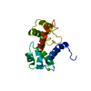
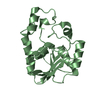
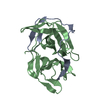
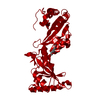
Mass: 24004.711 Da / Num. of mol.: 1
Source method: isolated from a genetically manipulated source
Details: Genomic DNA from the protease deficient ER1 strain derived form Thermobifida fusca YX was a gift from David B. Wilson at Cornell University.
Source: (gene. exp.) Thermobifida fusca (bacteria) / Strain: YX / Gene: Tfu_2693 / Plasmid: SpeedET / Production host: Escherichia Coli (E. coli) / Strain (production host): HK100 / References: UniProt: Q47LE6
#2: Chemical ChemComp-CO / COBALT (II) ION


Mass: 58.933 Da / Num. of mol.: 1 / Source method: obtained synthetically / Formula: Co
#3: Chemical ChemComp-HEZ / HEXANE-1,6-DIOL


Mass: 118.174 Da / Num. of mol.: 1 / Source method: obtained synthetically / Formula: C6H14O2
#4: Water ChemComp-HOH / water


Mass: 18.015 Da / Num. of mol.: 6 / Source method: isolated from a natural source / Formula: H2O
Sequence detailsTHE CONSTRUCT WAS EXPRESSED WITH A PURIFICATION TAG MGSDKIHHHHHHENLYFQG. THE TAG WAS REMOVED WITH ...THE CONSTRUCT WAS EXPRESSED WITH A PURIFICATION TAG MGSDKIHHHHHHENLYFQG. THE TAG WAS REMOVED WITH TEV PROTEASE LEAVING ONLY A GLYCINE (0) FOLLOWED BY RESIDUES 1-212 OF THE FULL LENGTH (252 AMINO ACID) TARGET SEQUENCE.

-
Experimental details

-
Experiment

ExperimentMethod: X-RAY DIFFRACTION / Number of used crystals: 1

-
Sample preparation

CrystalDensity Matthews: 2.85 Å3/Da / Density % sol: 56.89 %
Crystal growTemperature: 277 K / Method: vapor diffusion, sitting drop / pH: 4.6
Details: 1.0000M 1,6-Hexanediol, 0.0100M CoCl2, 0.1M Acetate pH 4.6, NANODROP, VAPOR DIFFUSION, SITTING DROP, temperature 277K

-
Data collection

DiffractionMean temperature: 100 K
Diffraction sourceSource: SYNCHROTRON / Site: SSRL / Beamline: BL11-1 / Wavelength: 0.97922
DetectorType: MARMOSAIC 325 mm CCD / Detector: CCD / Date: Apr 10, 2008 / Details: Flat mirror (vertical focusing)
RadiationMonochromator: Single crystal Si(111) bent monochromator (horizontal focusing)
Protocol: SINGLE WAVELENGTH / Monochromatic (M) / Laue (L): M / Scattering type: x-ray
Radiation wavelengthWavelength: 0.97922 Å / Relative weight: 1
ReflectionResolution: 2.65→48.97 Å / Num. obs: 8611 / % possible obs: 99.8 % / Observed criterion σ(I): -3 / Redundancy: 6.95 % / Biso Wilson estimate: 82.093 Å2 / Rmerge(I) obs: 0.048 / Net I/σ(I): 25.89
Reflection shell
Resolution (Å)Rmerge(I) obsMean I/σ(I) obsNum. measured obsNum. unique obsDiffraction-ID% possible all
2.65-2.740.812.156877941100
2.74-2.850.4533.860428431100
2.85-2.980.3274.959608361100
2.98-3.140.2167.861458641100
3.14-3.340.13213.260838581100
3.34-3.590.07222.258008261100
3.59-3.950.04931.95999853199.6
3.95-4.520.0348.260168701100
4.52-5.670.02655.15973883199.9
5.67-48.970.02260.76108986199.1

-
Phasing

PhasingMethod: molecular replacement

-
Processing

Software
NameVersionClassificationNB
REFMAC5.5.0102refinement
PHENIXrefinement
MolProbity3beta29model building
XSCALEdata scaling
PDB_EXTRACT3.006data extraction
XDSdata reduction
MOLREPphasing
RefinementMethod to determine structure: molecular replacement
Starting model: 2fug
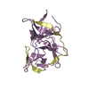
Resolution: 2.65→48.97 Å / Cor.coef. Fo:Fc: 0.952 / Cor.coef. Fo:Fc free: 0.923 / Occupancy max: 1 / Occupancy min: 0.5 / SU B: 22.045 / SU ML: 0.206 / TLS residual ADP flag: LIKELY RESIDUAL / Cross valid method: THROUGHOUT / σ(F): 0 / ESU R: 0.352 / ESU R Free: 0.273 / Stereochemistry target values: MAXIMUM LIKELIHOOD
Details: 1.HYDROGENS HAVE BEEN ADDED IN THE RIDING POSITIONS. 2.ATOM RECORD CONTAINS RESIDUAL B FACTORS ONLY 3.COBALT (CO) AND 1,6-HEXANEDIOL (HEZ) FROM THE CRYSTALLIZATION SOLUTION WERE MODELED INTO ...Details: 1.HYDROGENS HAVE BEEN ADDED IN THE RIDING POSITIONS. 2.ATOM RECORD CONTAINS RESIDUAL B FACTORS ONLY 3.COBALT (CO) AND 1,6-HEXANEDIOL (HEZ) FROM THE CRYSTALLIZATION SOLUTION WERE MODELED INTO THE STRUCTURE. THE MODELING OF COBALT IS SUPPORTED BY ANOMALOUS DIFFERENCE MAPS. 4. THE ELECTRON DENSITY CORRESPONDING TO THE N-TERMINAL 59 RESIDUES WAS DISORDERED AND THIS REGION COULD NOT BE MODELED.
RfactorNum. reflection% reflectionSelection details
Rfree0.259 402 4.7 %RANDOM
Rwork0.207 ---
obs0.21 8568 99.88 %-
Solvent computationIon probe radii: 0.8 Å / Shrinkage radii: 0.8 Å / VDW probe radii: 1.2 Å / Solvent model: MASK
Displacement parametersBiso max: 74.72 Å2 / Biso mean: 40.196 Å2 / Biso min: 19.26 Å2
Baniso -1Baniso -2Baniso -3
1-1.75 Å20 Å20 Å2
2--1.75 Å20 Å2
3----3.51 Å2
Refinement stepCycle: LAST / Resolution: 2.65→48.97 Å
ProteinNucleic acidLigandSolventTotal
Num. atoms1222 0 9 6 1237
Refine LS restraints
Refine-IDTypeDev idealDev ideal targetNumber
X-RAY DIFFRACTIONr_bond_refined_d0.0090.0211283
X-RAY DIFFRACTIONr_bond_other_d0.0020.02853
X-RAY DIFFRACTIONr_angle_refined_deg1.411.9471758
X-RAY DIFFRACTIONr_angle_other_deg0.96732061
X-RAY DIFFRACTIONr_dihedral_angle_1_deg2.9875157
X-RAY DIFFRACTIONr_dihedral_angle_2_deg29.20923.23568
X-RAY DIFFRACTIONr_dihedral_angle_3_deg11.55815179
X-RAY DIFFRACTIONr_dihedral_angle_4_deg11.8951511
X-RAY DIFFRACTIONr_chiral_restr0.0710.2191
X-RAY DIFFRACTIONr_gen_planes_refined0.0060.0211457
X-RAY DIFFRACTIONr_gen_planes_other0.0010.02276
X-RAY DIFFRACTIONr_mcbond_it1.0873780
X-RAY DIFFRACTIONr_mcbond_other0.1743307
X-RAY DIFFRACTIONr_mcangle_it2.12651269
X-RAY DIFFRACTIONr_scbond_it3.6058503
X-RAY DIFFRACTIONr_scangle_it5.75411487
LS refinement shellResolution: 2.65→2.719 Å / Total num. of bins used: 20
RfactorNum. reflection% reflection
Rfree0.435 35 -
Rwork0.371 585 -
all-620 -
obs--100 %
Refinement TLS params.Method: refined / Origin x: 53.536 Å / Origin y: 41.0403 Å / Origin z: 31.0986 Å
111213212223313233
T0.3335 Å20.0136 Å2-0.004 Å2-0.2358 Å2-0.0439 Å2--0.2211 Å2
L2.9653 °21.0644 °20.7181 °2-1.907 °21.2701 °2--2.7202 °2
S-0.0534 Å °-0.0611 Å °0.2599 Å °-0.1365 Å °0.0924 Å °-0.0751 Å °-0.1782 Å °0.1546 Å °-0.039 Å °

+
About Yorodumi

-
News

-
Feb 9, 2022. New format data for meta-information of EMDB entries

New format data for meta-information of EMDB entries

  • Version 3 of the EMDB header file is now the official format.
  • The previous official version 1.9 will be removed from the archive.

Related info.:EMDB header

External links:wwPDB to switch to version 3 of the EMDB data model

-
Aug 12, 2020. Covid-19 info

Covid-19 info

URL: https://pdbjlvh1.pdbj.org/emnavi/covid19.php

New page: Covid-19 featured information page in EM Navigator.

Related info.:Covid-19 info / Mar 5, 2020. Novel coronavirus structure data

+
Mar 5, 2020. Novel coronavirus structure data

Novel coronavirus structure data

Related info.:Yorodumi Speices / Aug 12, 2020. Covid-19 info

External links:COVID-19 featured content - PDBj / Molecule of the Month (242):Coronavirus Proteases

+
Jan 31, 2019. EMDB accession codes are about to change! (news from PDBe EMDB page)

EMDB accession codes are about to change! (news from PDBe EMDB page)

  • The allocation of 4 digits for EMDB accession codes will soon come to an end. Whilst these codes will remain in use, new EMDB accession codes will include an additional digit and will expand incrementally as the available range of codes is exhausted. The current 4-digit format prefixed with “EMD-” (i.e. EMD-XXXX) will advance to a 5-digit format (i.e. EMD-XXXXX), and so on. It is currently estimated that the 4-digit codes will be depleted around Spring 2019, at which point the 5-digit format will come into force.
  • The EM Navigator/Yorodumi systems omit the EMD- prefix.

Related info.:Q: What is EMD? / ID/Accession-code notation in Yorodumi/EM Navigator

External links:EMDB Accession Codes are Changing Soon! / Contact to PDBj

+
Jul 12, 2017. Major update of PDB

Major update of PDB

  • wwPDB released updated PDB data conforming to the new PDBx/mmCIF dictionary.
  • This is a major update changing the version number from 4 to 5, and with Remediation, in which all the entries are updated.
  • In this update, many items about electron microscopy experimental information are reorganized (e.g. em_software).
  • Now, EM Navigator and Yorodumi are based on the updated data.

External links:wwPDB Remediation / Enriched Model Files Conforming to OneDep Data Standards Now Available in the PDB FTP Archive

-
Yorodumi

Thousand views of thousand structures

  • Yorodumi is a browser for structure data from EMDB, PDB, SASBDB, etc.
  • This page is also the successor to EM Navigator detail page, and also detail information page/front-end page for Omokage search.
  • The word "yorodu" (or yorozu) is an old Japanese word meaning "ten thousand". "mi" (miru) is to see.

Related info.:EMDB / PDB / SASBDB / Comparison of 3 databanks / Yorodumi Search / Aug 31, 2016. New EM Navigator & Yorodumi / Yorodumi Papers / Jmol/JSmol / Function and homology information / Changes in new EM Navigator and Yorodumi

Read more