 Movie
Movie Controller

[English] 日本語
 Yorodumi
- PDB-3mb5: Crystal structure of P. abyssi tRNA m1A58 methyltransferase in co... -
+ Open data

- Basic information
| Entry | Database: PDB / ID: 3mb5 | ||||||
|---|---|---|---|---|---|---|---|
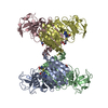
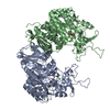
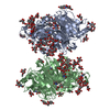
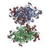
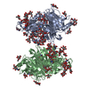
| Title | Crystal structure of P. abyssi tRNA m1A58 methyltransferase in complex with S-adenosyl-L-methionine | ||||||
 Components Components | SAM-dependent methyltransferase | ||||||
 Keywords Keywords | TRANSFERASE / RNA methyltransferase / m1A / TrmI / intermolecular contacts / region-specificity / tetramer / disulfide bond / hyperthermostability / Methyltransferase | ||||||
| Function / homology |  Function and homology information Function and homology informationtRNA (adenine57-N1/adenine58-N1)-methyltransferase / tRNA (adenine(57)-N1)/(adenine(58)-N1)-methyltransferase activity / tRNA (m1A) methyltransferase complex / tRNA (adenine(58)-N1)-methyltransferase activity / tRNA methylation Similarity search - Function | ||||||
| Biological species |   Pyrococcus abyssi (archaea) Pyrococcus abyssi (archaea) | ||||||
| Method |  X-RAY DIFFRACTION / SYNCHROTRON / MOLECULAR REPLACEMENT / Resolution: 1.6 Å X-RAY DIFFRACTION / SYNCHROTRON / MOLECULAR REPLACEMENT / Resolution: 1.6 Å | ||||||
 Authors Authors | Guelorget, A. / Golinelli-Pimpaneau, B. | ||||||
 Citation Citation | Journal: Nucleic Acids Res. / Year: 2010 Title: Insights into the hyperthermostability and unusual region-specificity of archaeal Pyrococcus abyssi tRNA m1A57/58 methyltransferase. Authors: Guelorget, A. / Roovers, M. / Guerineau, V. / Barbey, C. / Li, X. / Golinelli-Pimpaneau, B. #1: Journal: Nucleic Acids Res. / Year: 2004 Title: A primordial RNA modification enzyme: the case of tRNA (m1A) methyltransferase Authors: Roovers, M. / Wouters, J. / Bujnicki, J.M. / Tricot, C. / Stalon, V. / Grosjean, H. / Droogmans, L. | ||||||
| History |
|
- Structure visualization
| Structure viewer | Molecule: MolmilJmol/JSmol |
|---|
- Downloads & links
-Download
| PDBx/mmCIF format | 3mb5.cif.gz | 76.2 KB | Display | PDBx/mmCIF format |
|---|---|---|---|---|
| PDB format | pdb3mb5.ent.gz | 55.4 KB | Display | PDB format |
| PDBx/mmJSON format | 3mb5.json.gz | Tree view | PDBx/mmJSON format | |
| Others |  Other downloads Other downloads |
-Validation report
| Arichive directory | https://data.pdbj.org/pub/pdb/validation_reports/mb/3mb5ftp://data.pdbj.org/pub/pdb/validation_reports/mb/3mb5 | HTTPS FTP |
|---|
-Related structure data
| Related structure data | 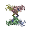 3lgaSC  3lhdC S: Starting model for refinement C: citing same article ( |
|---|---|
| Similar structure data |


-Links
 PDBj
PDBj
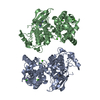
- Assembly
| Deposited unit | 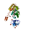
| ||||||||
|---|---|---|---|---|---|---|---|---|---|
| 1 | 
| ||||||||
| Unit cell |
| ||||||||
| Components on special symmetry positions |
|
-Components
-Protein , 1 types, 1 molecules A
| #1: Protein | Mass: 29162.932 Da / Num. of mol.: 1 Source method: isolated from a genetically manipulated source Source: (gene. exp.) Pyrococcus abyssi (archaea) / Strain: GE5 / Gene: PAB0283, pimT-like, PYRAB04300 / Plasmid: pET30 / Production host:  |
|---|
-Non-polymers , 5 types, 352 molecules 


| #2: Chemical | ChemComp-SAM /  Mass: 398.437 Da / Num. of mol.: 1 / Source method: obtained synthetically / Formula: C15H22N6O5S Mass: 398.437 Da / Num. of mol.: 1 / Source method: obtained synthetically / Formula: C15H22N6O5S | ||||||
|---|---|---|---|---|---|---|---|
| #3: Chemical | ChemComp-SO4 /  Mass: 96.063 Da / Num. of mol.: 4 / Source method: obtained synthetically / Formula: SO4 Mass: 96.063 Da / Num. of mol.: 4 / Source method: obtained synthetically / Formula: SO4#4: Chemical | ChemComp-EDO /  Mass: 62.068 Da / Num. of mol.: 6 / Source method: obtained synthetically / Formula: C2H6O2 Mass: 62.068 Da / Num. of mol.: 6 / Source method: obtained synthetically / Formula: C2H6O2#5: Chemical | ChemComp-ACT / |  Mass: 59.044 Da / Num. of mol.: 1 / Source method: obtained synthetically / Formula: C2H3O2 Mass: 59.044 Da / Num. of mol.: 1 / Source method: obtained synthetically / Formula: C2H3O2#6: Water | ChemComp-HOH / | Mass: 18.015 Da / Num. of mol.: 340 / Source method: isolated from a natural source / Formula: H2O |
-Details
| Has protein modification | Y |
|---|
-Experimental details
-Experiment
| Experiment | Method: X-RAY DIFFRACTION / Number of used crystals: 1 |
|---|
- Sample preparation
| Crystal | Density Matthews: 3.65 Å3/Da / Density % sol: 66.33 % |
|---|---|
| Crystal grow | Temperature: 291 K / Method: vapor diffusion, hanging drop Details: 2.8M ammonium sulfate, 0.2M ammonium acetate, VAPOR DIFFUSION, HANGING DROP, temperature 291K |
-Data collection
| Diffraction | Mean temperature: 100 K |
|---|---|
| Diffraction source | Source: SYNCHROTRON / Site: SOLEIL  / Beamline: PROXIMA 1 / Wavelength: 0.98 Å / Beamline: PROXIMA 1 / Wavelength: 0.98 Å |
| Detector | Type: ADSC QUANTUM 315r / Detector: CCD / Date: Feb 3, 2010 / Details: bi-morph mirrors |
| Radiation | Monochromator: cryogenically cooled monochromator crystal / Protocol: SINGLE WAVELENGTH / Monochromatic (M) / Laue (L): M / Scattering type: x-ray |
| Radiation wavelength | Wavelength: 0.98 Å / Relative weight: 1 |
| Reflection | Resolution: 1.6→20 Å / Num. obs: 56562 / % possible obs: 99.8 % / Redundancy: 9.1 % / Biso Wilson estimate: 22 Å2 / Rmerge(I) obs: 0.103 / Net I/σ(I): 16.42 |
| Reflection shell | Resolution: 1.6→1.7 Å / Redundancy: 5.8 % / Rmerge(I) obs: 0.317 / Mean I/σ(I) obs: 5.1 / Num. unique all: 9295 / % possible all: 99.9 |
- Processing
| Software |
| |||||||||||||||||||||||||||||||||||||||||||||||||||||||||||||||||
|---|---|---|---|---|---|---|---|---|---|---|---|---|---|---|---|---|---|---|---|---|---|---|---|---|---|---|---|---|---|---|---|---|---|---|---|---|---|---|---|---|---|---|---|---|---|---|---|---|---|---|---|---|---|---|---|---|---|---|---|---|---|---|---|---|---|---|
| Refinement | Method to determine structure: MOLECULAR REPLACEMENT Starting model: PDB code 3LGA Resolution: 1.6→19.7 Å / Cor.coef. Fo:Fc: 0.966 / Cor.coef. Fo:Fc free: 0.96 / SU B: 1.298 / SU ML: 0.046 / Cross valid method: THROUGHOUT / ESU R: 0.071 / ESU R Free: 0.069 / Stereochemistry target values: MAXIMUM LIKELIHOOD / Details: HYDROGENS HAVE BEEN ADDED IN THE RIDING POSITIONS
| |||||||||||||||||||||||||||||||||||||||||||||||||||||||||||||||||
| Solvent computation | Ion probe radii: 0.8 Å / Shrinkage radii: 0.8 Å / VDW probe radii: 1.4 Å / Solvent model: MASK | |||||||||||||||||||||||||||||||||||||||||||||||||||||||||||||||||
| Displacement parameters | Biso mean: 25.151 Å2
| |||||||||||||||||||||||||||||||||||||||||||||||||||||||||||||||||
| Refinement step | Cycle: LAST / Resolution: 1.6→19.7 Å
| |||||||||||||||||||||||||||||||||||||||||||||||||||||||||||||||||
| Refine LS restraints |
| |||||||||||||||||||||||||||||||||||||||||||||||||||||||||||||||||
| LS refinement shell | Resolution: 1.6→1.641 Å / Total num. of bins used: 20
|
