 Movie
Movie Controller

[English] 日本語
 Yorodumi
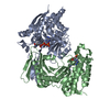
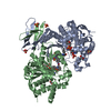
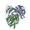
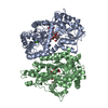
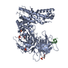
- PDB-3mar: Crystal structure of homodimeric R132H mutant of human cytosolic ... -
+ Open data

- Basic information
| Entry | Database: PDB / ID: 3mar | ||||||
|---|---|---|---|---|---|---|---|
| Title | Crystal structure of homodimeric R132H mutant of human cytosolic NADP(+)-dependent isocitrate dehydrogenase in complex with NADP | ||||||
 Components Components | Isocitrate dehydrogenase [NADP] cytoplasmic | ||||||
 Keywords Keywords | OXIDOREDUCTASE / isocitrate dehydrogenase / nicotinamide adenine dinucleotide phosphate / Rossmann fold | ||||||
| Function / homology |  Function and homology information Function and homology informationAbnormal conversion of 2-oxoglutarate to 2-hydroxyglutarate / NADPH regeneration / regulation of phospholipid catabolic process / regulation of phospholipid biosynthetic process / NFE2L2 regulating TCA cycle genes / isocitrate metabolic process / isocitrate dehydrogenase (NADP+) / NADPH regeneration / isocitrate dehydrogenase (NADP+) activity / NADP+ metabolic process ...Abnormal conversion of 2-oxoglutarate to 2-hydroxyglutarate / NADPH regeneration / regulation of phospholipid catabolic process / regulation of phospholipid biosynthetic process / NFE2L2 regulating TCA cycle genes / isocitrate metabolic process / isocitrate dehydrogenase (NADP+) / NADPH regeneration / isocitrate dehydrogenase (NADP+) activity / NADP+ metabolic process / 2-oxoglutarate metabolic process / glyoxylate cycle / response to steroid hormone / female gonad development / peroxisomal matrix / tricarboxylic acid cycle / glutathione metabolic process / Peroxisomal protein import / NAD binding / NADP binding / tertiary granule lumen / peroxisome / response to oxidative stress / secretory granule lumen / ficolin-1-rich granule lumen / cadherin binding / Neutrophil degranulation / magnesium ion binding / protein homodimerization activity / mitochondrion / extracellular exosome / extracellular region / identical protein binding / cytoplasm / cytosol Similarity search - Function | ||||||
| Biological species |  Homo sapiens (human) Homo sapiens (human) | ||||||
| Method |  X-RAY DIFFRACTION / SYNCHROTRON / MOLECULAR REPLACEMENT / Resolution: 3.41 Å X-RAY DIFFRACTION / SYNCHROTRON / MOLECULAR REPLACEMENT / Resolution: 3.41 Å | ||||||
 Authors Authors | Yang, B. / Peng, Y. / Ding, J. | ||||||
 Citation Citation | Journal: Cell Res. / Year: 2010 Title: Molecular mechanisms of "off-on switch" of activities of human IDH1 by tumor-associated mutation R132H. Authors: Yang, B. / Zhong, C. / Peng, Y. / Lai, Z. / Ding, J. | ||||||
| History |
|
- Structure visualization
| Structure viewer | Molecule: MolmilJmol/JSmol |
|---|
- Downloads & links
-Download
| PDBx/mmCIF format | 3mar.cif.gz | 155 KB | Display | PDBx/mmCIF format |
|---|---|---|---|---|
| PDB format | pdb3mar.ent.gz | 124.4 KB | Display | PDB format |
| PDBx/mmJSON format | 3mar.json.gz | Tree view | PDBx/mmJSON format | |
| Others |  Other downloads Other downloads |
-Validation report
| Arichive directory | https://data.pdbj.org/pub/pdb/validation_reports/ma/3marftp://data.pdbj.org/pub/pdb/validation_reports/ma/3mar | HTTPS FTP |
|---|
-Related structure data
| Related structure data | 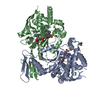 3mapC 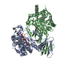 3masC 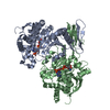 1t09S C: citing same article ( S: Starting model for refinement |
|---|---|
| Similar structure data |


-Links
 PDBj
PDBj

- Assembly
| Deposited unit | 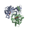
| ||||||||
|---|---|---|---|---|---|---|---|---|---|
| 1 |
| ||||||||
| Unit cell |
|
-Components
| #1: Protein | Mass: 47772.316 Da / Num. of mol.: 2 / Mutation: R132H Source method: isolated from a genetically manipulated source Source: (gene. exp.) Homo sapiens (human) / Gene: IDH1, PICD / Plasmid: pRSF-Duet / Production host:  References: UniProt: O75874, isocitrate dehydrogenase (NADP+) #2: Chemical |   Mass: 743.405 Da / Num. of mol.: 2 / Source method: obtained synthetically / Formula: C21H28N7O17P3 Mass: 743.405 Da / Num. of mol.: 2 / Source method: obtained synthetically / Formula: C21H28N7O17P3 |
|---|
-Experimental details
-Experiment
| Experiment | Method: X-RAY DIFFRACTION / Number of used crystals: 1 |
|---|
- Sample preparation
| Crystal | Density Matthews: 2.83 Å3/Da / Density % sol: 56.52 % |
|---|---|
| Crystal grow | Temperature: 277 K / Method: vapor diffusion, hanging drop / pH: 8.2 Details: 1.0M Na2HPO4/KH2PO4, pH 8.2, vapor diffusion, hanging drop, temperature 277K |
-Data collection
| Diffraction | Mean temperature: 100 K |
|---|---|
| Diffraction source | Source: SYNCHROTRON / Site: Photon Factory  / Beamline: AR-NW12A / Wavelength: 1 Å / Beamline: AR-NW12A / Wavelength: 1 Å |
| Detector | Type: ADSC QUANTUM 210 / Detector: CCD / Date: Mar 30, 2009 |
| Radiation | Protocol: SINGLE WAVELENGTH / Monochromatic (M) / Laue (L): M / Scattering type: x-ray |
| Radiation wavelength | Wavelength: 1 Å / Relative weight: 1 |
| Reflection | Resolution: 3.4→50 Å / Num. obs: 14215 / % possible obs: 91.2 % / Redundancy: 4.4 % / Rmerge(I) obs: 0.183 / Net I/σ(I): 5.2 |
| Reflection shell | Resolution: 3.4→3.52 Å / Redundancy: 3.9 % / Rmerge(I) obs: 0.561 / % possible all: 87.6 |
- Processing
| Software |
| ||||||||||||||||||||||||||||||||||||||||||||||||||||||||||||||||||||||||||||||||
|---|---|---|---|---|---|---|---|---|---|---|---|---|---|---|---|---|---|---|---|---|---|---|---|---|---|---|---|---|---|---|---|---|---|---|---|---|---|---|---|---|---|---|---|---|---|---|---|---|---|---|---|---|---|---|---|---|---|---|---|---|---|---|---|---|---|---|---|---|---|---|---|---|---|---|---|---|---|---|---|---|---|
| Refinement | Method to determine structure: MOLECULAR REPLACEMENT Starting model: PDB ENTRY 1T09 Resolution: 3.41→50 Å / SU B: 36.264 / SU ML: 0.553 / Cross valid method: THROUGHOUT / σ(F): 0 / ESU R Free: 0.746 / Stereochemistry target values: MAXIMUM LIKELIHOOD / Details: U VALUES : REFINED INDIVIDUALLY
| ||||||||||||||||||||||||||||||||||||||||||||||||||||||||||||||||||||||||||||||||
| Solvent computation | Ion probe radii: 0.8 Å / Shrinkage radii: 0.8 Å / VDW probe radii: 1.4 Å / Solvent model: MASK | ||||||||||||||||||||||||||||||||||||||||||||||||||||||||||||||||||||||||||||||||
| Displacement parameters | Biso mean: 89.2 Å2
| ||||||||||||||||||||||||||||||||||||||||||||||||||||||||||||||||||||||||||||||||
| Refinement step | Cycle: LAST / Resolution: 3.41→50 Å
| ||||||||||||||||||||||||||||||||||||||||||||||||||||||||||||||||||||||||||||||||
| Refine LS restraints |
| ||||||||||||||||||||||||||||||||||||||||||||||||||||||||||||||||||||||||||||||||
| LS refinement shell | Resolution: 3.4→3.52 Å / Total num. of bins used: 10
|
