 Movie
Movie Controller

[English] 日本語
 Yorodumi
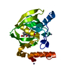
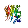
- PDB-3m6r: Crystal structure of Arabidopsis thaliana peptide deformylase 1B ... -
+ Open data

- Basic information
| Entry | Database: PDB / ID: 3m6r | ||||||
|---|---|---|---|---|---|---|---|
| Title | Crystal structure of Arabidopsis thaliana peptide deformylase 1B (AtPDF1B) G41M mutant in complex with actinonin | ||||||
 Components Components | Peptide deformylase 1B | ||||||
 Keywords Keywords | HYDROLASE/ANTIBIOTIC / peptide deformylase / 1B / PDF / N-terminal excision pathway / NME / Arabidopsis thaliana / induced-fit / Hydrolase / Metal-binding / Mitochondrion / Protein biosynthesis / Transit peptide / HYDROLASE-ANTIBIOTIC complex | ||||||
| Function / homology |  Function and homology information Function and homology informationpeptide deformylase / peptide deformylase activity / chloroplast stroma / plastid / chloroplast / translation / mitochondrion / metal ion binding Similarity search - Function | ||||||
| Biological species |  | ||||||
| Method |  X-RAY DIFFRACTION / SYNCHROTRON / MOLECULAR REPLACEMENT / Resolution: 2.4 Å X-RAY DIFFRACTION / SYNCHROTRON / MOLECULAR REPLACEMENT / Resolution: 2.4 Å | ||||||
 Authors Authors | Fieulaine, S. / Meinnel, T. / Giglione, C. | ||||||
 Citation Citation | Journal: Plos Biol. / Year: 2011 Title: Trapping conformational states along ligand-binding dynamics of peptide deformylase: the impact of induced fit on enzyme catalysis Authors: Fieulaine, S. / Boularot, A. / Artaud, I. / Desmadril, M. / Dardel, F. / Meinnel, T. / Giglione, C. | ||||||
| History |
|
- Structure visualization
| Structure viewer | Molecule: MolmilJmol/JSmol |
|---|
- Downloads & links
-Download
| PDBx/mmCIF format | 3m6r.cif.gz | 168.3 KB | Display | PDBx/mmCIF format |
|---|---|---|---|---|
| PDB format | pdb3m6r.ent.gz | 131.3 KB | Display | PDB format |
| PDBx/mmJSON format | 3m6r.json.gz | Tree view | PDBx/mmJSON format | |
| Others |  Other downloads Other downloads |
-Validation report
| Arichive directory | https://data.pdbj.org/pub/pdb/validation_reports/m6/3m6rftp://data.pdbj.org/pub/pdb/validation_reports/m6/3m6r | HTTPS FTP |
|---|
-Related structure data
| Related structure data | 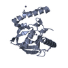 3m6oSC 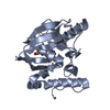 3m6pC 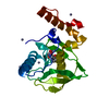 3m6qC 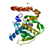 3o3jC 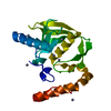 3pn2C  3pn3C  3pn4C  3pn5C 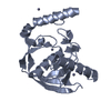 3pn6C S: Starting model for refinement C: citing same article ( |
|---|---|
| Similar structure data |


-Links
 PDBj
PDBj- Assembly
| Deposited unit | 
| ||||||||
|---|---|---|---|---|---|---|---|---|---|
| 1 | 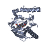
| ||||||||
| 2 | 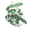
| ||||||||
| 3 | 
| ||||||||
| 4 | 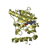
| ||||||||
| Unit cell |
|
-Components
| #1: Protein | Mass: 22050.377 Da / Num. of mol.: 4 / Fragment: residues 1-193 / Mutation: G41M Source method: isolated from a genetically manipulated source Source: (gene. exp.)  #2: Chemical | ChemComp-BB2 /   Mass: 385.498 Da / Num. of mol.: 4 / Source method: obtained synthetically / Formula: C19H35N3O5 / Comment: antitumor, antibiotic*YM Mass: 385.498 Da / Num. of mol.: 4 / Source method: obtained synthetically / Formula: C19H35N3O5 / Comment: antitumor, antibiotic*YM#3: Chemical | ChemComp-ZN /   Mass: 65.409 Da / Num. of mol.: 19 / Source method: obtained synthetically / Formula: Zn Mass: 65.409 Da / Num. of mol.: 19 / Source method: obtained synthetically / Formula: Zn#4: Water | ChemComp-HOH / |  Mass: 18.015 Da / Num. of mol.: 464 / Source method: isolated from a natural source / Formula: H2O Mass: 18.015 Da / Num. of mol.: 464 / Source method: isolated from a natural source / Formula: H2O |
|---|
-Experimental details
-Experiment
| Experiment | Method: X-RAY DIFFRACTION / Number of used crystals: 1 |
|---|
- Sample preparation
| Crystal | Density Matthews: 2.39 Å3/Da / Density % sol: 48.55 % |
|---|---|
| Crystal grow | Temperature: 293 K / Method: vapor diffusion, hanging drop Details: 14% PEG-3350, 0.1M zinc acetate, VAPOR DIFFUSION, HANGING DROP, temperature 293K |
-Data collection
| Diffraction | Mean temperature: 100 K |
|---|---|
| Diffraction source | Source: SYNCHROTRON / Site: ESRF  / Beamline: ID23-2 / Wavelength: 0.873 Å / Beamline: ID23-2 / Wavelength: 0.873 Å |
| Detector | Type: MARMOSAIC 225 mm CCD / Detector: CCD / Date: Jul 25, 2008 |
| Radiation | Protocol: SINGLE WAVELENGTH / Monochromatic (M) / Laue (L): M / Scattering type: x-ray |
| Radiation wavelength | Wavelength: 0.873 Å / Relative weight: 1 |
| Reflection | Resolution: 2.4→50 Å / Num. all: 33981 / Num. obs: 33143 / % possible obs: 97.5 % / Observed criterion σ(F): 2 / Observed criterion σ(I): 2 / Redundancy: 6 % / Biso Wilson estimate: 37.8 Å2 / Rsym value: 0.057 / Net I/σ(I): 21.28 |
| Reflection shell | Resolution: 2.4→2.54 Å / Redundancy: 6 % / Mean I/σ(I) obs: 4.69 / Num. unique all: 5176 / Rsym value: 0.42 / % possible all: 96.1 |
- Processing
| Software |
| ||||||||||||||||||||||||||||||||||||||||||||||||||||||||||||
|---|---|---|---|---|---|---|---|---|---|---|---|---|---|---|---|---|---|---|---|---|---|---|---|---|---|---|---|---|---|---|---|---|---|---|---|---|---|---|---|---|---|---|---|---|---|---|---|---|---|---|---|---|---|---|---|---|---|---|---|---|---|
| Refinement | Method to determine structure: MOLECULAR REPLACEMENT Starting model: PDB entry 3M6O Resolution: 2.4→47.15 Å / Rfactor Rfree error: 0.006 / Data cutoff high absF: 1872314.99 / Data cutoff low absF: 0 / Isotropic thermal model: RESTRAINED / Cross valid method: THROUGHOUT / Stereochemistry target values: MAXIMUM LIKELIHOOD
| ||||||||||||||||||||||||||||||||||||||||||||||||||||||||||||
| Solvent computation | Solvent model: FLAT MODEL / Bsol: 50.538 Å2 / ksol: 0.319753 e/Å3 | ||||||||||||||||||||||||||||||||||||||||||||||||||||||||||||
| Displacement parameters | Biso mean: 46 Å2
| ||||||||||||||||||||||||||||||||||||||||||||||||||||||||||||
| Refine analyze |
| ||||||||||||||||||||||||||||||||||||||||||||||||||||||||||||
| Refinement step | Cycle: LAST / Resolution: 2.4→47.15 Å
| ||||||||||||||||||||||||||||||||||||||||||||||||||||||||||||
| Refine LS restraints |
| ||||||||||||||||||||||||||||||||||||||||||||||||||||||||||||
| LS refinement shell | Resolution: 2.4→2.55 Å / Rfactor Rfree error: 0.018 / Total num. of bins used: 6
| ||||||||||||||||||||||||||||||||||||||||||||||||||||||||||||
| Xplor file |
|
