 Movie
Movie Controller

[English] 日本語
 Yorodumi
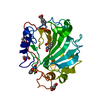
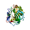
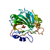
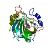
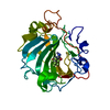
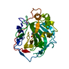
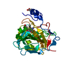
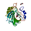
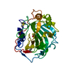
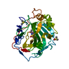
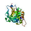
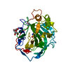
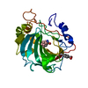
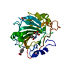
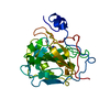
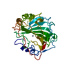
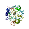
- PDB-3m14: Carbonic Anhydrase II in complex with novel sulfonamide inhibitor -
+ Open data

- Basic information
| Entry | Database: PDB / ID: 3m14 | ||||||
|---|---|---|---|---|---|---|---|
| Title | Carbonic Anhydrase II in complex with novel sulfonamide inhibitor | ||||||
 Components Components | Carbonic anhydrase 2 | ||||||
 Keywords Keywords | LYASE/LYASE INHIBITOR / 10 Stranded Twisted Beta-Sheets / Lyase / Disease mutation / Metal-binding / LYASE-LYASE INHIBITOR complex | ||||||
| Function / homology |  Function and homology information Function and homology information: / positive regulation of dipeptide transmembrane transport / secretion / regulation of monoatomic anion transport / cyanamide hydratase / cyanamide hydratase activity / arylesterase activity / regulation of chloride transport / Reversible hydration of carbon dioxide / positive regulation of synaptic transmission, GABAergic ...: / positive regulation of dipeptide transmembrane transport / secretion / regulation of monoatomic anion transport / cyanamide hydratase / cyanamide hydratase activity / arylesterase activity / regulation of chloride transport / Reversible hydration of carbon dioxide / positive regulation of synaptic transmission, GABAergic / morphogenesis of an epithelium / angiotensin-activated signaling pathway / Developmental Lineage of Pancreatic Ductal Cells / carbonic anhydrase / regulation of intracellular pH / carbonate dehydratase activity / neuron cellular homeostasis / carbon dioxide transport / Erythrocytes take up oxygen and release carbon dioxide / Erythrocytes take up carbon dioxide and release oxygen / apical part of cell / myelin sheath / extracellular exosome / zinc ion binding / plasma membrane / cytosol / cytoplasm Similarity search - Function | ||||||
| Biological species |  Homo sapiens (human) Homo sapiens (human) | ||||||
| Method |  X-RAY DIFFRACTION / SYNCHROTRON / FOURIER SYNTHESIS / Resolution: 1.38 Å X-RAY DIFFRACTION / SYNCHROTRON / FOURIER SYNTHESIS / Resolution: 1.38 Å | ||||||
 Authors Authors | Schulze Wischeler, J. / Heine, A. / Klebe, G. / Sandner, N.U. | ||||||
 Citation Citation | Journal: To be Published Title: Structural investigation and inhibitor studies on Carbonic Anhydrase II Authors: Schulze Wischeler, J. / Sandner, N.U. / Haake, M. / Supuran, C. / Heine, A. / Klebe, G. | ||||||
| History |
|
- Structure visualization
| Structure viewer | Molecule: MolmilJmol/JSmol |
|---|
- Downloads & links
-Download
| PDBx/mmCIF format | 3m14.cif.gz | 130.2 KB | Display | PDBx/mmCIF format |
|---|---|---|---|---|
| PDB format | pdb3m14.ent.gz | 99.7 KB | Display | PDB format |
| PDBx/mmJSON format | 3m14.json.gz | Tree view | PDBx/mmJSON format | |
| Others |  Other downloads Other downloads |
-Validation report
| Arichive directory | https://data.pdbj.org/pub/pdb/validation_reports/m1/3m14ftp://data.pdbj.org/pub/pdb/validation_reports/m1/3m14 | HTTPS FTP |
|---|
-Related structure data
| Related structure data |  3m04C  3m2xC 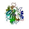 1oq5S C: citing same article ( S: Starting model for refinement |
|---|---|
| Similar structure data |


-Links
 PDBj
PDBj

- Assembly
| Deposited unit | 
| ||||||||
|---|---|---|---|---|---|---|---|---|---|
| 1 |
| ||||||||
| Unit cell |
|
-Components
| #1: Protein | Mass: 29806.588 Da / Num. of mol.: 1 / Fragment: Carbonic Anhydrase II Source method: isolated from a genetically manipulated source Source: (gene. exp.) Homo sapiens (human) / Gene: CA2 / Plasmid: pGEX-4T1 / Production host:  |
|---|---|
| #2: Chemical | ChemComp-ZN /   Mass: 65.409 Da / Num. of mol.: 1 / Source method: obtained synthetically / Formula: Zn Mass: 65.409 Da / Num. of mol.: 1 / Source method: obtained synthetically / Formula: Zn |
| #3: Chemical | ChemComp-BEV /   Mass: 181.237 Da / Num. of mol.: 1 / Source method: obtained synthetically / Formula: C3H7N3O2S2 Mass: 181.237 Da / Num. of mol.: 1 / Source method: obtained synthetically / Formula: C3H7N3O2S2 |
| #4: Chemical | ChemComp-BE7 / (  Mass: 357.156 Da / Num. of mol.: 1 / Source method: obtained synthetically / Formula: C7H5ClHgO2 / Comment: protease inhibitor*YM Mass: 357.156 Da / Num. of mol.: 1 / Source method: obtained synthetically / Formula: C7H5ClHgO2 / Comment: protease inhibitor*YM |
| #5: Water | ChemComp-HOH /  Mass: 18.015 Da / Num. of mol.: 255 / Source method: isolated from a natural source / Formula: H2O Mass: 18.015 Da / Num. of mol.: 255 / Source method: isolated from a natural source / Formula: H2O |
-Experimental details
-Experiment
| Experiment | Method: X-RAY DIFFRACTION / Number of used crystals: 1 |
|---|
- Sample preparation
| Crystal | Density Matthews: 2.15 Å3/Da / Density % sol: 39.71 % |
|---|---|
| Crystal grow | Temperature: 291 K / Method: vapor diffusion, sitting drop / pH: 7.8 Details: 2.5 M Ammoniumsulfate 50 mM Tris 0.1 mM p-chloromercuribenzoic acid 1 mM Sulfonamide, pH 7.8, VAPOR DIFFUSION, SITTING DROP, temperature 291K |
-Data collection
| Diffraction | Mean temperature: 110 K |
|---|---|
| Diffraction source | Source: SYNCHROTRON / Site: SLS  / Beamline: X06DA / Wavelength: 1 Å / Beamline: X06DA / Wavelength: 1 Å |
| Detector | Type: MARMOSAIC 225 mm CCD / Detector: CCD / Date: Aug 1, 2009 |
| Radiation | Monochromator: Bartels monochromator / Protocol: SINGLE WAVELENGTH / Monochromatic (M) / Laue (L): M / Scattering type: x-ray |
| Radiation wavelength | Wavelength: 1 Å / Relative weight: 1 |
| Reflection | Resolution: 1.38→25 Å / Num. all: 49221 / Num. obs: 49221 / % possible obs: 99.1 % / Observed criterion σ(F): 0 / Observed criterion σ(I): 0 / Redundancy: 3.6 % / Rsym value: 0.064 / Net I/σ(I): 13.7 |
| Reflection shell | Resolution: 1.38→1.4 Å / Redundancy: 2.6 % / Mean I/σ(I) obs: 5.5 / Num. unique all: 2281 / Rsym value: 0.162 / % possible all: 91.6 |
- Processing
| Software |
| |||||||||||||||||||||||||||||||||
|---|---|---|---|---|---|---|---|---|---|---|---|---|---|---|---|---|---|---|---|---|---|---|---|---|---|---|---|---|---|---|---|---|---|---|
| Refinement | Method to determine structure: FOURIER SYNTHESIS Starting model: 1OQ5 Resolution: 1.38→10 Å / Num. parameters: 21269 / Num. restraintsaints: 26986 / Cross valid method: FREE R / σ(F): 0 / Stereochemistry target values: ENGH AND HUBER Details: ANISOTROPIC REFINEMENT REDUCED FREE R (NO CUTOFF) BY The Method of Parkin, Moezzi & Hope,J.appl.cryst.28(1995)53-56 Anisotropic refinement reduced free r (no cutoff)
| |||||||||||||||||||||||||||||||||
| Refine analyze | Num. disordered residues: 17 / Occupancy sum hydrogen: 1981 / Occupancy sum non hydrogen: 2311.8 | |||||||||||||||||||||||||||||||||
| Refinement step | Cycle: LAST / Resolution: 1.38→10 Å
| |||||||||||||||||||||||||||||||||
| Refine LS restraints |
|
