 Movie
Movie Controller

[English] 日本語
 Yorodumi
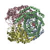
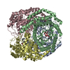
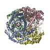
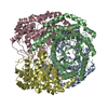
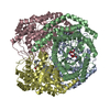
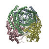
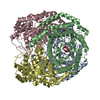
- PDB-3m0h: Crystal structure of Pseudomonas stutzeri L-rhamnose isomerase mu... -
+ Open data

- Basic information
| Entry | Database: PDB / ID: 3m0h | ||||||
|---|---|---|---|---|---|---|---|
| Title | Crystal structure of Pseudomonas stutzeri L-rhamnose isomerase mutant S329F in complex with L-rhamnose | ||||||
 Components Components | L-rhamnose isomerase | ||||||
 Keywords Keywords | ISOMERASE / BETA/ALPHA BARREL / HOMO-TETRAMER / METAL-BINDING PROTEIN / TIM barrel | ||||||
| Function / homology |  Function and homology information Function and homology information | ||||||
| Biological species |  Pseudomonas stutzeri (bacteria) Pseudomonas stutzeri (bacteria) | ||||||
| Method |  X-RAY DIFFRACTION / SYNCHROTRON / MOLECULAR REPLACEMENT / Resolution: 1.58 Å X-RAY DIFFRACTION / SYNCHROTRON / MOLECULAR REPLACEMENT / Resolution: 1.58 Å | ||||||
 Authors Authors | Yoshida, H. / Takeda, K. / Izumori, K. / Kamitori, S. | ||||||
 Citation Citation | Journal: Protein Eng.Des.Sel. / Year: 2010 Title: Elucidation of the role of Ser329 and the C-terminal region in the catalytic activity of Pseudomonas stutzeri L-rhamnose isomerase Authors: Yoshida, H. / Takeda, K. / Izumori, K. / Kamitori, S. #1: Journal: J.Mol.Biol. / Year: 2007Title: The structures of L-rhamnose isomerase from Pseudomonas stutzeri in complexes with L-rhamnose and D-allose provide insights into broad substrate specificity Authors: Yoshida, H. / Yamada, M. / Ohyama, Y. / Takada, G. / Izumori, K. / Kamitori, S. #2: Journal: Febs J. / Year: 2010Title: Catalytic reaction mechanism of Pseudomonas stutzeri L-rhamnose isomerase deduced from X-ray structures Authors: Yoshida, H. / Yamaji, M. / Ishii, T. / Izumori, K. / Kamitori, S. | ||||||
| History |
|
- Structure visualization
| Structure viewer | Molecule: MolmilJmol/JSmol |
|---|
- Downloads & links
-Download
| PDBx/mmCIF format | 3m0h.cif.gz | 365.5 KB | Display | PDBx/mmCIF format |
|---|---|---|---|---|
| PDB format | pdb3m0h.ent.gz | 292.8 KB | Display | PDB format |
| PDBx/mmJSON format | 3m0h.json.gz | Tree view | PDBx/mmJSON format | |
| Others |  Other downloads Other downloads |
-Validation report
| Arichive directory | https://data.pdbj.org/pub/pdb/validation_reports/m0/3m0hftp://data.pdbj.org/pub/pdb/validation_reports/m0/3m0h | HTTPS FTP |
|---|
-Related structure data
| Related structure data | 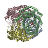 3m0lC 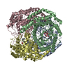 3m0mC  3m0vC  3m0xC 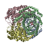 3m0yC 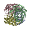 2hcvS S: Starting model for refinement C: citing same article ( |
|---|---|
| Similar structure data |


-Links
 PDBj
PDBj
- Assembly
| Deposited unit | 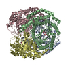
| ||||||||
|---|---|---|---|---|---|---|---|---|---|
| 1 |
| ||||||||
| Unit cell |
|
-Components
| #1: Protein | Mass: 48064.707 Da / Num. of mol.: 4 / Mutation: D150N, S329F Source method: isolated from a genetically manipulated source Source: (gene. exp.) Pseudomonas stutzeri (bacteria) / Plasmid: pQE60 / Production host: #2: Chemical | ChemComp-MN /   Mass: 54.938 Da / Num. of mol.: 8 / Source method: obtained synthetically / Formula: Mn Mass: 54.938 Da / Num. of mol.: 8 / Source method: obtained synthetically / Formula: Mn#3: Sugar | ChemComp-RNS / 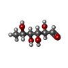  Type: L-saccharide / Mass: 164.156 Da / Num. of mol.: 4 Type: L-saccharide / Mass: 164.156 Da / Num. of mol.: 4Source method: isolated from a genetically manipulated source Formula: C6H12O5 #4: Water | ChemComp-HOH / |  Mass: 18.015 Da / Num. of mol.: 1674 / Source method: isolated from a natural source / Formula: H2O Mass: 18.015 Da / Num. of mol.: 1674 / Source method: isolated from a natural source / Formula: H2O |
|---|
-Experimental details
-Experiment
| Experiment | Method: X-RAY DIFFRACTION / Number of used crystals: 1 |
|---|
- Sample preparation
| Crystal | Density Matthews: 2.09 Å3/Da / Density % sol: 41.16 % |
|---|---|
| Crystal grow | Temperature: 293 K / Method: vapor diffusion, hanging drop / pH: 6.3 Details: 7-8% (w/v) polyethylene glycol 20000, 50mM MES pH 6.3, VAPOR DIFFUSION, HANGING DROP, temperature 293K |
-Data collection
| Diffraction | Mean temperature: 100 K |
|---|---|
| Diffraction source | Source: SYNCHROTRON / Site: Photon Factory  / Beamline: BL-5A / Wavelength: 1 Å / Beamline: BL-5A / Wavelength: 1 Å |
| Detector | Type: ADSC QUANTUM 315 / Detector: CCD / Date: Jun 12, 2007 |
| Radiation | Monochromator: GRAPHITE / Protocol: SINGLE WAVELENGTH / Monochromatic (M) / Laue (L): M / Scattering type: x-ray |
| Radiation wavelength | Wavelength: 1 Å / Relative weight: 1 |
| Reflection | Resolution: 1.58→50 Å / Num. all: 211383 / Num. obs: 211383 / % possible obs: 97.9 % / Observed criterion σ(F): 0 / Observed criterion σ(I): 0 / Redundancy: 3.6 % / Biso Wilson estimate: 14.6 Å2 / Rmerge(I) obs: 0.053 / Net I/σ(I): 13.9 |
| Reflection shell | Resolution: 1.58→1.64 Å / Redundancy: 3.3 % / Rmerge(I) obs: 0.212 / Mean I/σ(I) obs: 4.9 / % possible all: 94.4 |
- Processing
| Software |
| |||||||||||||||||||||||||
|---|---|---|---|---|---|---|---|---|---|---|---|---|---|---|---|---|---|---|---|---|---|---|---|---|---|---|
| Refinement | Method to determine structure: MOLECULAR REPLACEMENT Starting model: PDB ENTRY 2HCV Resolution: 1.58→32.71 Å / Rfactor Rfree error: 0.001 / Data cutoff high absF: 82493.25 / Data cutoff low absF: 0 / Isotropic thermal model: RESTRAINED / Cross valid method: THROUGHOUT / σ(F): 0 / Stereochemistry target values: Engh & Huber
| |||||||||||||||||||||||||
| Solvent computation | Solvent model: FLAT MODEL / Bsol: 46.4321 Å2 / ksol: 0.359028 e/Å3 | |||||||||||||||||||||||||
| Displacement parameters | Biso mean: 13.5 Å2
| |||||||||||||||||||||||||
| Refine analyze |
| |||||||||||||||||||||||||
| Refinement step | Cycle: LAST / Resolution: 1.58→32.71 Å
| |||||||||||||||||||||||||
| Refine LS restraints |
| |||||||||||||||||||||||||
| LS refinement shell | Resolution: 1.58→1.68 Å / Rfactor Rfree error: 0.004 / Total num. of bins used: 6
| |||||||||||||||||||||||||
| Xplor file |
|
