 Movie
Movie Controller

[English] 日本語
 Yorodumi
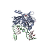
- PDB-3m05: The crystal structure of a functionally unknown protein PEPE_1480... -
+ Open data

- Basic information
| Entry | Database: PDB / ID: 3m05 | ||||||
|---|---|---|---|---|---|---|---|
| Title | The crystal structure of a functionally unknown protein PEPE_1480 from Pediococcus pentosaceus ATCC 25745 | ||||||
 Components Components | uncharacterized protein PEPE_1480 | ||||||
 Keywords Keywords | structural genomics / unknown function / PSI-2 / protein structure initiative / midwest center for structural genomics / MCSG | ||||||
| Function / homology | Cyclic-di-AMP receptor / Cyclic-di-AMP receptor / Alpha-Beta Plaits - #120 / Nitrogen regulatory PII-like, alpha/beta / Nitrogen regulatory protein PII/ATP phosphoribosyltransferase, C-terminal / Alpha-Beta Plaits / 2-Layer Sandwich / Alpha Beta / Protein from nitrogen regulatory protein P-II (GLNB) family Function and homology information Function and homology information | ||||||
| Biological species |  Pediococcus pentosaceus (bacteria) Pediococcus pentosaceus (bacteria) | ||||||
| Method |  X-RAY DIFFRACTION / SYNCHROTRON / MAD / Resolution: 3.145 Å X-RAY DIFFRACTION / SYNCHROTRON / MAD / Resolution: 3.145 Å | ||||||
 Authors Authors | Tan, K. / Bigelow, L. / Bearden, J. / Joachimiak, A. / Midwest Center for Structural Genomics (MCSG) | ||||||
 Citation Citation | Journal: To be Published Title: The crystal structure of a functionally unknown protein PEPE_1480 from Pediococcus pentosaceus ATCC 25745 Authors: Tan, K. / Bigelow, L. / Bearden, J. / Joachimiak, A. | ||||||
| History |
|
- Structure visualization
| Structure viewer | Molecule: MolmilJmol/JSmol |
|---|
- Downloads & links
-Download
| PDBx/mmCIF format | 3m05.cif.gz | 129.7 KB | Display | PDBx/mmCIF format |
|---|---|---|---|---|
| PDB format | pdb3m05.ent.gz | 104.5 KB | Display | PDB format |
| PDBx/mmJSON format | 3m05.json.gz | Tree view | PDBx/mmJSON format | |
| Others |  Other downloads Other downloads |
-Validation report
| Summary document | 3m05_validation.pdf.gz | 458 KB | Display | wwPDB validaton report |
|---|---|---|---|---|
| Full document | 3m05_full_validation.pdf.gz | 468.8 KB | Display | |
| Data in XML | 3m05_validation.xml.gz | 14.9 KB | Display | |
| Data in CIF | 3m05_validation.cif.gz | 18.4 KB | Display | |
| Arichive directory | https://data.pdbj.org/pub/pdb/validation_reports/m0/3m05ftp://data.pdbj.org/pub/pdb/validation_reports/m0/3m05 | HTTPS FTP |
-Related structure data
| Similar structure data | |
|---|---|
| Other databases |


-Links
 PDBj
PDBj- Assembly
| Deposited unit | 
| ||||||||
|---|---|---|---|---|---|---|---|---|---|
| 1 |
| ||||||||
| 2 | 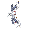
| ||||||||
| 3 | 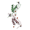
| ||||||||
| Unit cell |
| ||||||||
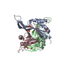
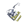
| Details | Experimentally unknown. The chains A, B and C may form a trimer. |
-Components
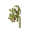
| #1: Protein | Mass: 12843.794 Da / Num. of mol.: 3 Source method: isolated from a genetically manipulated source Source: (gene. exp.) Pediococcus pentosaceus (bacteria) / Strain: ATCC 25745 / Gene: PEPE_1480 / Plasmid: pMCSG19 / Production host: #2: Chemical | ChemComp-SO4 /   Mass: 96.063 Da / Num. of mol.: 6 / Source method: obtained synthetically / Formula: SO4 Mass: 96.063 Da / Num. of mol.: 6 / Source method: obtained synthetically / Formula: SO4#3: Water | ChemComp-HOH / |  Mass: 18.015 Da / Num. of mol.: 5 / Source method: isolated from a natural source / Formula: H2O Mass: 18.015 Da / Num. of mol.: 5 / Source method: isolated from a natural source / Formula: H2OHas protein modification | Y | |
|---|
-Experimental details
-Experiment
| Experiment | Method: X-RAY DIFFRACTION / Number of used crystals: 1 |
|---|
- Sample preparation
| Crystal | Density Matthews: 5.55 Å3/Da / Density % sol: 77.86 % |
|---|---|
| Crystal grow | Temperature: 277 K / Method: vapor diffusion, sitting drop / pH: 6 Details: 0.2M Li2SO4, 0.1M MES, 20% (v/v)1,4-butanodiol, pH 6.0, VAPOR DIFFUSION, SITTING DROP, temperature 277K |
-Data collection
| Diffraction | Mean temperature: 100 K | |||||||||
|---|---|---|---|---|---|---|---|---|---|---|
| Diffraction source | Source: SYNCHROTRON / Site: APS  / Beamline: 19-ID / Wavelength: 0.97937, 0.97951 / Beamline: 19-ID / Wavelength: 0.97937, 0.97951 | |||||||||
| Detector | Type: ADSC QUANTUM 315r / Detector: CCD / Date: Feb 4, 2010 / Details: Mirror | |||||||||
| Radiation | Monochromator: Si 111 crystal / Protocol: MAD / Monochromatic (M) / Laue (L): M / Scattering type: x-ray | |||||||||
| Radiation wavelength |
| |||||||||
| Reflection | Resolution: 3.145→37.862 Å / Num. all: 15624 / Num. obs: 15624 / % possible obs: 99.7 % / Observed criterion σ(F): 0 / Observed criterion σ(I): 0 / Redundancy: 7 % / Rmerge(I) obs: 0.109 / Net I/σ(I): 27.2 | |||||||||
| Reflection shell | Resolution: 3.15→3.2 Å / Redundancy: 7.2 % / Rmerge(I) obs: 0.804 / Mean I/σ(I) obs: 2.4 / Num. unique all: 749 / % possible all: 100 |
- Processing
| Software |
| ||||||||||||||||||||||||||||||||||||||||||||||||||||||||||||||||||||||||||||||||||||||||||||||||||||||||
|---|---|---|---|---|---|---|---|---|---|---|---|---|---|---|---|---|---|---|---|---|---|---|---|---|---|---|---|---|---|---|---|---|---|---|---|---|---|---|---|---|---|---|---|---|---|---|---|---|---|---|---|---|---|---|---|---|---|---|---|---|---|---|---|---|---|---|---|---|---|---|---|---|---|---|---|---|---|---|---|---|---|---|---|---|---|---|---|---|---|---|---|---|---|---|---|---|---|---|---|---|---|---|---|---|---|
| Refinement | Method to determine structure: MAD / Resolution: 3.145→37.862 Å / SU ML: 0.26 / σ(F): 0.05 / Stereochemistry target values: ML
| ||||||||||||||||||||||||||||||||||||||||||||||||||||||||||||||||||||||||||||||||||||||||||||||||||||||||
| Solvent computation | Shrinkage radii: 0.9 Å / VDW probe radii: 1.11 Å / Solvent model: FLAT BULK SOLVENT MODEL / Bsol: 60.084 Å2 / ksol: 0.293 e/Å3 | ||||||||||||||||||||||||||||||||||||||||||||||||||||||||||||||||||||||||||||||||||||||||||||||||||||||||
| Refinement step | Cycle: LAST / Resolution: 3.145→37.862 Å
| ||||||||||||||||||||||||||||||||||||||||||||||||||||||||||||||||||||||||||||||||||||||||||||||||||||||||
| Refine LS restraints |
| ||||||||||||||||||||||||||||||||||||||||||||||||||||||||||||||||||||||||||||||||||||||||||||||||||||||||
| LS refinement shell |
| ||||||||||||||||||||||||||||||||||||||||||||||||||||||||||||||||||||||||||||||||||||||||||||||||||||||||
| Refinement TLS params. | Refine-ID: X-RAY DIFFRACTION
| ||||||||||||||||||||||||||||||||||||||||||||||||||||||||||||||||||||||||||||||||||||||||||||||||||||||||
| Refinement TLS group | Selection details: chain C |
