 Movie
Movie Controller


+ Open data

- Basic information
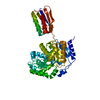
| Entry | Database: PDB / ID: 3lyq | ||||||
|---|---|---|---|---|---|---|---|
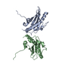
| Title | Crystal structure of IpgB2 from Shigella flexneri | ||||||
 Components Components | IpgB2 | ||||||
 Keywords Keywords | RHOA-BINDING PROTEIN / IpgB2 / GEF / WxxxE / TTSS effector protein / bacterial GEF / cytoskeleton dynamics | ||||||
| Function / homology | SopE-like GEF fold - #20 / Bacterial effector protein IpgB-like / IpaB/EvcA family / SopE-like GEF fold / Orthogonal Bundle / Mainly Alpha / MU-OXO-DIIRON / CITRATE ANION / IpgB2 Function and homology information Function and homology information | ||||||
| Biological species |  Shigella flexneri (bacteria) Shigella flexneri (bacteria) | ||||||
| Method |  X-RAY DIFFRACTION / SYNCHROTRON / SAD / Resolution: 2.3 Å X-RAY DIFFRACTION / SYNCHROTRON / SAD / Resolution: 2.3 Å | ||||||
 Authors Authors | Klink, B.U. / Barden, S. / Heidler, T.V. / Borchers, C. / Ladwein, M. / Stradal, T.E.B. / Rottner, K. / Heinz, D.W. | ||||||
 Citation Citation | Journal: J.Biol.Chem. / Year: 2010 Title: Structure of Shigella IPGB2 in complex with human RhoA: Implications for the mechanism of bacterial GEF-mimicry Authors: Klink, B.U. / Barden, S. / Heidler, T.V. / Borchers, C. / Ladwein, M. / Stradal, T.E.B. / Rottner, K. / Heinz, D.W. | ||||||
| History |
|
- Structure visualization
| Structure viewer | Molecule: MolmilJmol/JSmol |
|---|
- Downloads & links
-Download
| PDBx/mmCIF format | 3lyq.cif.gz | 90.2 KB | Display | PDBx/mmCIF format |
|---|---|---|---|---|
| PDB format | pdb3lyq.ent.gz | 69.6 KB | Display | PDB format |
| PDBx/mmJSON format | 3lyq.json.gz | Tree view | PDBx/mmJSON format | |
| Others |  Other downloads Other downloads |
-Validation report
| Arichive directory | https://data.pdbj.org/pub/pdb/validation_reports/ly/3lyqftp://data.pdbj.org/pub/pdb/validation_reports/ly/3lyq | HTTPS FTP |
|---|
-Related structure data


-Links
 PDBj
PDBj


- Assembly
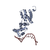
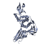
| Deposited unit | 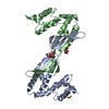
| ||||||||
|---|---|---|---|---|---|---|---|---|---|
| 1 | 
| ||||||||
| 2 | 
| ||||||||
| Unit cell |
| ||||||||
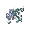
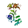
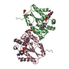
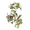
| Details | THE ASYMMETRIC UNIT CONTAINS TWO BIOLOGICAL UNITS, WHICH INTERACT VIA DOMAIN-SWAPPING. |
-Components
| #1: Protein | Mass: 21935.990 Da / Num. of mol.: 2 Source method: isolated from a genetically manipulated source Source: (gene. exp.) Shigella flexneri (bacteria) / Gene: ipgB2 / Plasmid: pET-M 41 / Production host: #2: Chemical |   Mass: 189.100 Da / Num. of mol.: 3 / Source method: obtained synthetically / Formula: C6H5O7 Mass: 189.100 Da / Num. of mol.: 3 / Source method: obtained synthetically / Formula: C6H5O7#3: Chemical |   Mass: 127.689 Da / Num. of mol.: 2 / Source method: obtained synthetically / Formula: Fe2O Mass: 127.689 Da / Num. of mol.: 2 / Source method: obtained synthetically / Formula: Fe2O#4: Water | ChemComp-HOH / |  Mass: 18.015 Da / Num. of mol.: 106 / Source method: isolated from a natural source / Formula: H2O Mass: 18.015 Da / Num. of mol.: 106 / Source method: isolated from a natural source / Formula: H2O |
|---|
-Experimental details
-Experiment
| Experiment | Method: X-RAY DIFFRACTION / Number of used crystals: 2 |
|---|
- Sample preparation
| Crystal | Density Matthews: 3.29 Å3/Da / Density % sol: 62.63 % |
|---|---|
| Crystal grow | Temperature: 277 K / Method: vapor diffusion, hanging drop / pH: 5.5 Details: 5% (w/w) PEG 3350, 7% isopropanol, 10mM FeCl3, 100mM trisodium citrate/citric acid pH 5.5, 10mM NaCl, vapor diffusion, hanging drop, temperature 277K |
-Data collection
| Diffraction |
| |||||||||||||||||||||||||||||||||||||||||||||||||||||||||||||||||||||||||||||
|---|---|---|---|---|---|---|---|---|---|---|---|---|---|---|---|---|---|---|---|---|---|---|---|---|---|---|---|---|---|---|---|---|---|---|---|---|---|---|---|---|---|---|---|---|---|---|---|---|---|---|---|---|---|---|---|---|---|---|---|---|---|---|---|---|---|---|---|---|---|---|---|---|---|---|---|---|---|---|
| Diffraction source |
| |||||||||||||||||||||||||||||||||||||||||||||||||||||||||||||||||||||||||||||
| Detector |
| |||||||||||||||||||||||||||||||||||||||||||||||||||||||||||||||||||||||||||||
| Radiation |
| |||||||||||||||||||||||||||||||||||||||||||||||||||||||||||||||||||||||||||||
| Radiation wavelength |
| |||||||||||||||||||||||||||||||||||||||||||||||||||||||||||||||||||||||||||||
| Reflection | Redundancy: 11.8 % / Av σ(I) over netI: 7.4 / Number: 186094 / Rmerge(I) obs: 0.07 / Rsym value: 0.07 / D res high: 2.8 Å / D res low: 90.909 Å / Num. obs: 15757 / % possible obs: 99.3 | |||||||||||||||||||||||||||||||||||||||||||||||||||||||||||||||||||||||||||||
| Diffraction reflection shell | ID: 1
| |||||||||||||||||||||||||||||||||||||||||||||||||||||||||||||||||||||||||||||
| Reflection | Resolution: 2.3→22.88 Å / Num. obs: 25363 / % possible obs: 95.4 % / Observed criterion σ(I): -3 / Redundancy: 18.2 % / Biso Wilson estimate: 49.2 Å2 / Rmerge(I) obs: 0.056 / Net I/σ(I): 33.35 | |||||||||||||||||||||||||||||||||||||||||||||||||||||||||||||||||||||||||||||
| Reflection shell | Resolution: 2.3→2.36 Å / Redundancy: 6.4 % / Rmerge(I) obs: 0.675 / Mean I/σ(I) obs: 2.6 / Num. measured obs: 7703 / Num. unique all: 1205 / Num. unique obs: 1205 / % possible all: 63 |

-Phasing
| Phasing | Method: SAD |
|---|
- Processing
| Software |
| |||||||||||||||||||||||||||||||||||||||||||||||||||||||||||||||||||||||||||||||||||||||||||||||||||||||||||||||||||||||||||||||||||||||||||||||||||||||||||||||||||||||||||||||
|---|---|---|---|---|---|---|---|---|---|---|---|---|---|---|---|---|---|---|---|---|---|---|---|---|---|---|---|---|---|---|---|---|---|---|---|---|---|---|---|---|---|---|---|---|---|---|---|---|---|---|---|---|---|---|---|---|---|---|---|---|---|---|---|---|---|---|---|---|---|---|---|---|---|---|---|---|---|---|---|---|---|---|---|---|---|---|---|---|---|---|---|---|---|---|---|---|---|---|---|---|---|---|---|---|---|---|---|---|---|---|---|---|---|---|---|---|---|---|---|---|---|---|---|---|---|---|---|---|---|---|---|---|---|---|---|---|---|---|---|---|---|---|---|---|---|---|---|---|---|---|---|---|---|---|---|---|---|---|---|---|---|---|---|---|---|---|---|---|---|---|---|---|---|---|---|---|
| Refinement | Method to determine structure: SAD / Resolution: 2.3→22.88 Å / Cor.coef. Fo:Fc: 0.946 / Cor.coef. Fo:Fc free: 0.913 / Occupancy max: 1 / Occupancy min: 0.5 / SU B: 17.362 / SU ML: 0.196 / TLS residual ADP flag: LIKELY RESIDUAL / Cross valid method: THROUGHOUT / σ(I): -3 / ESU R: 0.281 / ESU R Free: 0.249 / Stereochemistry target values: MAXIMUM LIKELIHOOD Details: HYDROGENS HAVE BEEN ADDED IN THE RIDING POSITIONS U VALUES : RESIDUAL ONLY; An almost spherical complex of ligands mediates a strong crystallographic contact between four different IpgB2 ...Details: HYDROGENS HAVE BEEN ADDED IN THE RIDING POSITIONS U VALUES : RESIDUAL ONLY; An almost spherical complex of ligands mediates a strong crystallographic contact between four different IpgB2 molecules. The cluster contains at least two iron atoms with a Fe-Fe-distance identical to the distance in MU-OXO-DIIRON. An interpretation as a Fe-citrate cluster containing four citrate molecules lowers both free and working R factor significantly. However, the precise architecture of the cluster used for this interpretation (chain A or B) only represents the "best compromise". Ligands in chain A or B may not be completely correct, contain unrealistic atom distances and do not perfectly describe the electron density.
| |||||||||||||||||||||||||||||||||||||||||||||||||||||||||||||||||||||||||||||||||||||||||||||||||||||||||||||||||||||||||||||||||||||||||||||||||||||||||||||||||||||||||||||||
| Solvent computation | Ion probe radii: 0.8 Å / Shrinkage radii: 0.8 Å / VDW probe radii: 1.4 Å / Solvent model: MASK | |||||||||||||||||||||||||||||||||||||||||||||||||||||||||||||||||||||||||||||||||||||||||||||||||||||||||||||||||||||||||||||||||||||||||||||||||||||||||||||||||||||||||||||||
| Displacement parameters | Biso max: 119.13 Å2 / Biso mean: 45.838 Å2 / Biso min: 10.85 Å2
| |||||||||||||||||||||||||||||||||||||||||||||||||||||||||||||||||||||||||||||||||||||||||||||||||||||||||||||||||||||||||||||||||||||||||||||||||||||||||||||||||||||||||||||||
| Refinement step | Cycle: LAST / Resolution: 2.3→22.88 Å
| |||||||||||||||||||||||||||||||||||||||||||||||||||||||||||||||||||||||||||||||||||||||||||||||||||||||||||||||||||||||||||||||||||||||||||||||||||||||||||||||||||||||||||||||
| Refine LS restraints |
| |||||||||||||||||||||||||||||||||||||||||||||||||||||||||||||||||||||||||||||||||||||||||||||||||||||||||||||||||||||||||||||||||||||||||||||||||||||||||||||||||||||||||||||||
| LS refinement shell | Resolution: 2.302→2.361 Å / Total num. of bins used: 20
| |||||||||||||||||||||||||||||||||||||||||||||||||||||||||||||||||||||||||||||||||||||||||||||||||||||||||||||||||||||||||||||||||||||||||||||||||||||||||||||||||||||||||||||||
| Refinement TLS params. | Method: refined / Refine-ID: X-RAY DIFFRACTION
| |||||||||||||||||||||||||||||||||||||||||||||||||||||||||||||||||||||||||||||||||||||||||||||||||||||||||||||||||||||||||||||||||||||||||||||||||||||||||||||||||||||||||||||||
| Refinement TLS group |
|
