heme oxygenase (staphylobilin-producing) / iron import into cell / heme oxygenase (decyclizing) activity / heme catabolic process / iron ion binding / heme binding / cytoplasm Similarity search - Function
Mass: 18.015 Da / Num. of mol.: 184 / Source method: isolated from a natural source / Formula: H2O
Nonpolymer details
MAGNESIUM ION 600 IS COORDINATED BY 6 WATERS 601-606.
-
Experimental details
-
Experiment
Experiment
Method: X-RAY DIFFRACTION / Number of used crystals: 1
-
Sample preparation
Crystal
Density Matthews: 2.68 Å3/Da / Density % sol: 54.15 % / Mosaicity: 1.09 °
Crystal grow
Temperature: 277 K / Method: vapor diffusion, sitting drop / pH: 5.5 Details: 25% polyethylene glycol 3350, 0.2M MgCl2, 0.1M Bis-Tris buffer pH 5.5, and soaked in 50mM sodium dithionite for 10 minutes with a cryoprotectant of 10% ethylene glycol before freezing, VAPOR ...Details: 25% polyethylene glycol 3350, 0.2M MgCl2, 0.1M Bis-Tris buffer pH 5.5, and soaked in 50mM sodium dithionite for 10 minutes with a cryoprotectant of 10% ethylene glycol before freezing, VAPOR DIFFUSION, SITTING DROP, temperature 277K
Type: MARMOSAIC 300 mm CCD / Detector: CCD / Date: Nov 8, 2008 Details: DCM with cryo-cooled 1st crystal sagittally bent 2nd crystal followed by vertically focusing mirror.
Radiation
Monochromator: double crystal monochromator (DCM) / Protocol: SINGLE WAVELENGTH / Monochromatic (M) / Laue (L): M / Scattering type: x-ray
In the structure databanks used in Yorodumi, some data are registered as the other names, "COVID-19 virus" and "2019-nCoV". Here are the details of the virus and the list of structure data.
Jan 31, 2019. EMDB accession codes are about to change! (news from PDBe EMDB page)
EMDB accession codes are about to change! (news from PDBe EMDB page)
The allocation of 4 digits for EMDB accession codes will soon come to an end. Whilst these codes will remain in use, new EMDB accession codes will include an additional digit and will expand incrementally as the available range of codes is exhausted. The current 4-digit format prefixed with “EMD-” (i.e. EMD-XXXX) will advance to a 5-digit format (i.e. EMD-XXXXX), and so on. It is currently estimated that the 4-digit codes will be depleted around Spring 2019, at which point the 5-digit format will come into force.
The EM Navigator/Yorodumi systems omit the EMD- prefix.
Related info.:Q: What is EMD? / ID/Accession-code notation in Yorodumi/EM Navigator
Yorodumi is a browser for structure data from EMDB, PDB, SASBDB, etc.
This page is also the successor to EM Navigator detail page, and also detail information page/front-end page for Omokage search.
The word "yorodu" (or yorozu) is an old Japanese word meaning "ten thousand". "mi" (miru) is to see.
Related info.:EMDB / PDB / SASBDB / Comparison of 3 databanks / Yorodumi Search / Aug 31, 2016. New EM Navigator & Yorodumi / Yorodumi Papers / Jmol/JSmol / Function and homology information / Changes in new EM Navigator and Yorodumi
 Movie
Movie Controller


 Open data
 Basic information
 Components
 Keywords
 Function and homology information
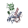
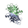
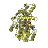
 Staphylococcus aureus (bacteria)
 X-RAY DIFFRACTION /
  Authors
 Citation
 Structure visualization
 Downloads & links
 Other downloads


 PDBj
PDBj Assembly
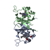

 Mass: 616.487 Da / Num. of mol.: 2 / Source method: obtained synthetically / Formula: C34H32FeN4O4
 Mass: 24.305 Da / Num. of mol.: 1 / Source method: obtained synthetically / Formula: Mg
 Mass: 18.015 Da / Num. of mol.: 184 / Source method: isolated from a natural source / Formula: H2O
 Sample preparation
 / Beamline: 08ID-1 / Wavelength: 0.97934 Å
 Processing