 Movie
Movie Controller

[English] 日本語
 Yorodumi
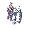
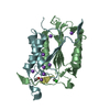
- PDB-3l2z: Crystal structure of hydrated Biotin Protein Ligase from M. tuber... -
+ Open data

- Basic information
| Entry | Database: PDB / ID: 3l2z | ||||||
|---|---|---|---|---|---|---|---|
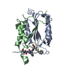
| Title | Crystal structure of hydrated Biotin Protein Ligase from M. tuberculosis | ||||||
 Components Components | BirA bifunctional protein | ||||||
 Keywords Keywords | LIGASE / BirA / hydrated structure / disordered ligand binding loops | ||||||
| Function / homology |  Function and homology information Function and homology informationbiotin-[biotin carboxyl-carrier protein] ligase / biotin--[biotin carboxyl-carrier protein] ligase activity / small molecule binding / post-translational protein modification / cytoplasm Similarity search - Function | ||||||
| Biological species |   Mycobacterium tuberculosis (bacteria) Mycobacterium tuberculosis (bacteria) | ||||||
| Method |  X-RAY DIFFRACTION / SYNCHROTRON / MOLECULAR REPLACEMENT / Resolution: 2.8 Å X-RAY DIFFRACTION / SYNCHROTRON / MOLECULAR REPLACEMENT / Resolution: 2.8 Å | ||||||
 Authors Authors | Gupta, V. / Gupta, R.K. / Khare, G. / Salunke, D.M. / Tyagi, A.K. | ||||||
 Citation Citation | Journal: Plos One / Year: 2010 Title: Structural ordering of disordered ligand-binding loops of biotin protein ligase into active conformations as a consequence of dehydration. Authors: Gupta, V. / Gupta, R.K. / Khare, G. / Salunke, D.M. / Surolia, A. / Tyagi, A.K. | ||||||
| History |
|
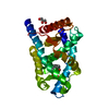
- Structure visualization
| Structure viewer | Molecule: MolmilJmol/JSmol |
|---|
- Downloads & links
-Download
| PDBx/mmCIF format | 3l2z.cif.gz | 99.8 KB | Display | PDBx/mmCIF format |
|---|---|---|---|---|
| PDB format | pdb3l2z.ent.gz | 76.7 KB | Display | PDB format |
| PDBx/mmJSON format | 3l2z.json.gz | Tree view | PDBx/mmJSON format | |
| Others |  Other downloads Other downloads |
-Validation report
| Arichive directory | https://data.pdbj.org/pub/pdb/validation_reports/l2/3l2zftp://data.pdbj.org/pub/pdb/validation_reports/l2/3l2z | HTTPS FTP |
|---|
-Related structure data
| Related structure data |  3l1aC  2cghS C: citing same article ( S: Starting model for refinement |
|---|---|
| Similar structure data |


-Links
 PDBj
PDBj
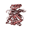
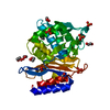
- Assembly
| Deposited unit | 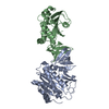
| ||||||||
|---|---|---|---|---|---|---|---|---|---|
| 1 | 
| ||||||||
| 2 | 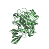
| ||||||||
| Unit cell |
|
-Components
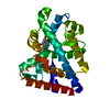
| #1: Protein | Mass: 28153.027 Da / Num. of mol.: 2 Source method: isolated from a genetically manipulated source Source: (gene. exp.) Mycobacterium tuberculosis (bacteria) / Strain: H37Rv / Gene: birA, MT3379, Rv3279c / Plasmid: pASK-IBA43plus / Production host: References: UniProt: P96884, biotin-[biotin carboxyl-carrier protein] ligase #2: Water | ChemComp-HOH / |  Mass: 18.015 Da / Num. of mol.: 26 / Source method: isolated from a natural source / Formula: H2O Mass: 18.015 Da / Num. of mol.: 26 / Source method: isolated from a natural source / Formula: H2O |
|---|
-Experimental details
-Experiment
| Experiment | Method: X-RAY DIFFRACTION / Number of used crystals: 4 |
|---|
- Sample preparation
| Crystal | Density Matthews: 2.35 Å3/Da / Density % sol: 47.63 % |
|---|---|
| Crystal grow | Temperature: 293 K / Method: vapor diffusion / pH: 7.5 Details: 12-16% PEG4000, PEG8000, 0.1M HEPES, pH 7.5, VAPOR DIFFUSION, temperature 293K |
-Data collection
| Diffraction | Mean temperature: 295 K |
|---|---|
| Diffraction source | Source: SYNCHROTRON / Site: EMBL/DESY, HAMBURG  / Beamline: X13 / Wavelength: 0.8088 Å / Beamline: X13 / Wavelength: 0.8088 Å |
| Detector | Type: MAR CCD 165 mm / Detector: CCD / Date: Feb 21, 2007 / Details: bending magnet |
| Radiation | Monochromator: Si 111, horizontally focussing / Protocol: SINGLE WAVELENGTH / Monochromatic (M) / Laue (L): M / Scattering type: x-ray |
| Radiation wavelength | Wavelength: 0.8088 Å / Relative weight: 1 |
| Reflection | Resolution: 2.8→15 Å / Num. obs: 15831 / % possible obs: 99.4 % / Observed criterion σ(F): 2 / Observed criterion σ(I): 2 / Redundancy: 5.7 % / Rmerge(I) obs: 0.098 / Net I/σ(I): 17.2 |
| Reflection shell | Resolution: 2.8→2.9 Å / Redundancy: 5.8 % / Rmerge(I) obs: 0.462 / Mean I/σ(I) obs: 2.2 / % possible all: 99.6 |
- Processing
| Software |
| ||||||||||||||||||||||||||||||||||||||||||||||||||||||||||||||||||
|---|---|---|---|---|---|---|---|---|---|---|---|---|---|---|---|---|---|---|---|---|---|---|---|---|---|---|---|---|---|---|---|---|---|---|---|---|---|---|---|---|---|---|---|---|---|---|---|---|---|---|---|---|---|---|---|---|---|---|---|---|---|---|---|---|---|---|---|
| Refinement | Method to determine structure: MOLECULAR REPLACEMENT Starting model: PDB entry 2CGH Resolution: 2.8→14.126 Å / SU ML: 0.66 / Cross valid method: THROUGHOUT / σ(F): 1.35 / Phase error: 23.61 / Stereochemistry target values: ML
| ||||||||||||||||||||||||||||||||||||||||||||||||||||||||||||||||||
| Solvent computation | Shrinkage radii: 0.9 Å / VDW probe radii: 1.11 Å / Solvent model: FLAT BULK SOLVENT MODEL / Bsol: 34.366 Å2 / ksol: 0.309 e/Å3 | ||||||||||||||||||||||||||||||||||||||||||||||||||||||||||||||||||
| Displacement parameters |
| ||||||||||||||||||||||||||||||||||||||||||||||||||||||||||||||||||
| Refinement step | Cycle: LAST / Resolution: 2.8→14.126 Å
| ||||||||||||||||||||||||||||||||||||||||||||||||||||||||||||||||||
| Refine LS restraints |
| ||||||||||||||||||||||||||||||||||||||||||||||||||||||||||||||||||
| LS refinement shell | Refine-ID: X-RAY DIFFRACTION / Total num. of bins used: 10
|
