Mass: 18.015 Da / Num. of mol.: 818 / Source method: isolated from a natural source / Formula: H2O
Has protein modification
Y
Sequence details
THE CONSTRUCT WAS EXPRESSED WITH A PURIFICATION TAG MGSDKIHHHHHHENLYFQG. THE TAG WAS REMOVED WITH ...THE CONSTRUCT WAS EXPRESSED WITH A PURIFICATION TAG MGSDKIHHHHHHENLYFQG. THE TAG WAS REMOVED WITH TEV PROTEASE LEAVING ONLY A GLYCINE (0) FOLLOWED BY THE TARGET SEQUENCE. THE CLONED CONSTRUCT CONTAINS RESIDUES 27-312 OF THE FULL LENGTH PROTEIN.
-
Experimental details
-
Experiment
Experiment
Method: X-RAY DIFFRACTION / Number of used crystals: 1
-
Sample preparation
Crystal
Density Matthews: 2.47 Å3/Da / Density % sol: 50.14 %
Crystal grow
Temperature: 277 K / Method: vapor diffusion, sitting drop / pH: 8.5 Details: 0.2000M MgCl2, 20.0000% PEG-8000, 0.1M TRIS pH 8.5, NANODROP, VAPOR DIFFUSION, SITTING DROP, temperature 277K
Type: MARMOSAIC 325 mm CCD / Detector: CCD / Date: Jun 11, 2009 / Details: Flat mirror (vertical focusing)
Radiation
Monochromator: Single crystal Si(111) bent monochromator (horizontal focusing) Protocol: MAD / Monochromatic (M) / Laue (L): M / Scattering type: x-ray
Radiation wavelength
ID
Wavelength (Å)
Relative weight
1
0.91837
1
2
0.9791
1
3
0.97858
1
Reflection
Resolution: 1.68→29.374 Å / Num. obs: 71903 / % possible obs: 99.6 % / Redundancy: 3.7 % / Biso Wilson estimate: 11.88 Å2 / Rmerge(I) obs: 0.132 / Rsym value: 0.132 / Net I/σ(I): 8.6
Reflection shell
Diffraction-ID: 1
Resolution (Å)
Redundancy (%)
Rmerge(I) obs
Mean I/σ(I) obs
Num. measured all
Num. unique all
Rsym value
% possible all
1.68-1.72
3.7
0.541
1.4
19446
5273
0.541
100
1.72-1.77
3.7
0.47
1.6
18869
5129
0.47
100
1.77-1.82
3.7
0.404
1.9
18445
4992
0.404
100
1.82-1.88
3.7
0.341
2.2
17938
4857
0.341
100
1.88-1.94
3.7
0.302
2.5
17502
4737
0.302
100
1.94-2.01
3.7
0.24
3
16879
4562
0.24
100
2.01-2.08
3.7
0.206
3.6
16416
4432
0.206
99.9
2.08-2.17
3.7
0.176
4.2
15754
4256
0.176
99.9
2.17-2.27
3.7
0.155
4.7
15099
4076
0.155
99.9
2.27-2.38
3.7
0.144
5
14579
3910
0.144
99.8
2.38-2.5
3.7
0.13
5.5
13769
3718
0.13
99.8
2.5-2.66
3.7
0.123
5.6
13142
3534
0.123
99.7
2.66-2.84
3.7
0.117
5.8
12232
3311
0.117
99.5
2.84-3.07
3.7
0.102
6.3
11408
3090
0.102
99.4
3.07-3.36
3.7
0.084
7.5
10515
2848
0.084
99.2
3.36-3.76
3.7
0.07
9
9488
2570
0.07
98.9
3.76-4.34
3.7
0.063
9.6
8442
2287
0.063
98.6
4.34-5.31
3.7
0.065
9.2
7182
1946
0.065
98.4
5.31-7.51
3.6
0.073
8.7
5528
1521
0.073
97.5
7.51-29.37
3.4
0.063
10
2943
854
0.063
94.6
-
Phasing
Phasing
Method: MAD
-
Processing
Software
Name
Version
Classification
NB
REFMAC
5.5.0102
refinement
PHENIX
refinement
SOLVE
phasing
MolProbity
3beta29
modelbuilding
SCALA
3.2.5
datascaling
PDB_EXTRACT
3.006
dataextraction
MOSFLM
datareduction
Refinement
Method to determine structure: MAD / Resolution: 1.68→29.374 Å / Cor.coef. Fo:Fc: 0.962 / Cor.coef. Fo:Fc free: 0.946 / Occupancy max: 1 / Occupancy min: 0.25 / SU B: 3.519 / SU ML: 0.053 / TLS residual ADP flag: LIKELY RESIDUAL / Cross valid method: THROUGHOUT / σ(F): 0 / ESU R: 0.084 / ESU R Free: 0.085 Stereochemistry target values: MAXIMUM LIKELIHOOD WITH PHASES Details: 1.HYDROGENS HAVE BEEN ADDED IN THE RIDING POSITIONS. 2.ATOM RECORD CONTAINS RESIDUAL B FACTORS ONLY. 3.A MET-INHIBITION PROTOCOL WAS USED FOR SELENOMETHIONINE INCORPORATION DURING PROTEIN ...Details: 1.HYDROGENS HAVE BEEN ADDED IN THE RIDING POSITIONS. 2.ATOM RECORD CONTAINS RESIDUAL B FACTORS ONLY. 3.A MET-INHIBITION PROTOCOL WAS USED FOR SELENOMETHIONINE INCORPORATION DURING PROTEIN EXPRESSION. THE OCCUPANCY OF THE SE ATOMS IN THE MSE RESIDUES WAS REDUCED TO 0.75 TO ACCOUNT FOR THE REDUCED SCATTERING POWER DUE TO PARTIAL S-MET INCORPORATION. 4.MAGNESIUM ATOMS (MG) AND POLYETHYLENE GLYCOL (PEG) FROM THE CRYSTALLIZATION SOLUTION ARE MODELED INTO THE STRUCTURE. 5. TLS PARAMETERS WERE ASSIGNED WITH THE AID OF THE TLS MOTION DETERMINATION SERVER. 6. A RAMACHANDRAN OUTLIER (A106) IS SUPPORTED BY CLEARLY DEFINED DENSITY.
Rfactor
Num. reflection
% reflection
Selection details
Rfree
0.182
3624
5 %
RANDOM
Rwork
0.15
-
-
-
obs
0.152
71861
99.41 %
-
Solvent computation
Ion probe radii: 0.8 Å / Shrinkage radii: 0.8 Å / VDW probe radii: 1.4 Å / Solvent model: MASK
In the structure databanks used in Yorodumi, some data are registered as the other names, "COVID-19 virus" and "2019-nCoV". Here are the details of the virus and the list of structure data.
Jan 31, 2019. EMDB accession codes are about to change! (news from PDBe EMDB page)
EMDB accession codes are about to change! (news from PDBe EMDB page)
The allocation of 4 digits for EMDB accession codes will soon come to an end. Whilst these codes will remain in use, new EMDB accession codes will include an additional digit and will expand incrementally as the available range of codes is exhausted. The current 4-digit format prefixed with “EMD-” (i.e. EMD-XXXX) will advance to a 5-digit format (i.e. EMD-XXXXX), and so on. It is currently estimated that the 4-digit codes will be depleted around Spring 2019, at which point the 5-digit format will come into force.
The EM Navigator/Yorodumi systems omit the EMD- prefix.
Related info.:Q: What is EMD? / ID/Accession-code notation in Yorodumi/EM Navigator
Yorodumi is a browser for structure data from EMDB, PDB, SASBDB, etc.
This page is also the successor to EM Navigator detail page, and also detail information page/front-end page for Omokage search.
The word "yorodu" (or yorozu) is an old Japanese word meaning "ten thousand". "mi" (miru) is to see.
Related info.:EMDB / PDB / SASBDB / Comparison of 3 databanks / Yorodumi Search / Aug 31, 2016. New EM Navigator & Yorodumi / Yorodumi Papers / Jmol/JSmol / Function and homology information / Changes in new EM Navigator and Yorodumi
 Movie
Movie Controller

 Yorodumi
 Open data
 Basic information
 Components
 Keywords
 Function and homology information
 Parabacteroides distasonis ATCC 8503 (bacteria)
 X-RAY DIFFRACTION /
  Authors
 Citation
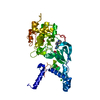
 Structure visualization
 Downloads & links
 Other downloads
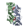


 PDBj
PDBj


 Assembly
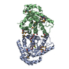

 Mass: 24.305 Da / Num. of mol.: 2 / Source method: obtained synthetically / Formula: Mg
 Mass: 106.120 Da / Num. of mol.: 3 / Source method: obtained synthetically / Formula: C4H10O3
 Mass: 18.015 Da / Num. of mol.: 818 / Source method: isolated from a natural source / Formula: H2O
 Sample preparation
 / Beamline: BL11-1 / Wavelength: 0.91837,0.97910,0.97858
 Processing