 Movie
Movie Controller

[English] 日本語
 Yorodumi
- PDB-3krp: Mint heterotetrameric geranyl pyrophosphate synthase in complex w... -
+ Open data

- Basic information
| Entry | Database: PDB / ID: 3krp | ||||||
|---|---|---|---|---|---|---|---|
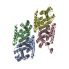
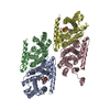
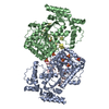
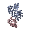
| Title | Mint heterotetrameric geranyl pyrophosphate synthase in complex with magnesium and GPP | ||||||
 Components Components | (Geranyl diphosphate synthase ...) x 2 | ||||||
 Keywords Keywords | TRANSFERASE / prenyltransferase / Isoprene biosynthesis / isoprenyl pyrophosphate synthase | ||||||
| Function / homology |  Function and homology information Function and homology informationgeranyl diphosphate biosynthetic process / prenyltransferase activity / geranylgeranyl diphosphate synthase activity / dimethylallyltranstransferase activity / metal ion binding / cytoplasm Similarity search - Function | ||||||
| Biological species |  Mentha x piperita (peppermint) Mentha x piperita (peppermint) | ||||||
| Method |  X-RAY DIFFRACTION / SYNCHROTRON / MOLECULAR REPLACEMENT / Resolution: 2.42 Å X-RAY DIFFRACTION / SYNCHROTRON / MOLECULAR REPLACEMENT / Resolution: 2.42 Å | ||||||
 Authors Authors | Chang, T.-H. / Hsieh, F.-L. / Ko, T.-P. / Wang, A.H.-J. | ||||||
 Citation Citation | Journal: Plant Cell / Year: 2010 Title: Structure of a heterotetrameric geranyl pyrophosphate synthase from mint (Mentha piperita) reveals intersubunit regulation Authors: Chang, T.-H. / Hsieh, F.-L. / Ko, T.-P. / Teng, K.-H. / Liang, P.-H. / Wang, A.H.-J. | ||||||
| History |
|
- Structure visualization
| Structure viewer | Molecule: MolmilJmol/JSmol |
|---|
- Downloads & links
-Download
| PDBx/mmCIF format | 3krp.cif.gz | 234 KB | Display | PDBx/mmCIF format |
|---|---|---|---|---|
| PDB format | pdb3krp.ent.gz | 184.6 KB | Display | PDB format |
| PDBx/mmJSON format | 3krp.json.gz | Tree view | PDBx/mmJSON format | |
| Others |  Other downloads Other downloads |
-Validation report
| Arichive directory | https://data.pdbj.org/pub/pdb/validation_reports/kr/3krpftp://data.pdbj.org/pub/pdb/validation_reports/kr/3krp | HTTPS FTP |
|---|
-Related structure data
| Related structure data |  3kraC  3krcC 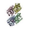 3krfC 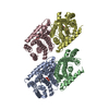 3kroC  2j1oS C: citing same article ( S: Starting model for refinement |
|---|---|
| Similar structure data |


-Links
 PDBj
PDBj
- Assembly
| Deposited unit | 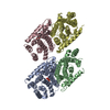
| ||||||||
|---|---|---|---|---|---|---|---|---|---|
| 1 |
| ||||||||
| 2 | 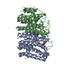
| ||||||||
| 3 | 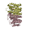
| ||||||||
| Unit cell |
| ||||||||
| Details | Gel filtration chromatography demonstrate that the biological assembly should be a hetero-tetramer, composed of two hetero-dimers (one is A chain and B chain, the other is C chain and D chain). |
-Components
-Geranyl diphosphate synthase ... , 2 types, 4 molecules ADBC
| #1: Protein | Mass: 31947.004 Da / Num. of mol.: 2 / Fragment: UNP residues 84-377 Source method: isolated from a genetically manipulated source Source: (gene. exp.) Mentha x piperita (peppermint) / Gene: GPPS Large Subunit / Plasmid: pET32 / Production host:  #2: Protein | Mass: 29755.963 Da / Num. of mol.: 2 / Fragment: UNP residues 49-313 Source method: isolated from a genetically manipulated source Source: (gene. exp.) Mentha x piperita (peppermint) / Gene: GPPS Small subunit / Plasmid: pET37 / Production host: |
|---|
-Non-polymers , 5 types, 823 molecules 


| #3: Chemical | ChemComp-PPV /  Mass: 177.975 Da / Num. of mol.: 1 / Source method: obtained synthetically / Formula: H4O7P2 Mass: 177.975 Da / Num. of mol.: 1 / Source method: obtained synthetically / Formula: H4O7P2 | ||||||
|---|---|---|---|---|---|---|---|
| #4: Chemical | ChemComp-MG /  Mass: 24.305 Da / Num. of mol.: 4 / Source method: obtained synthetically / Formula: Mg Mass: 24.305 Da / Num. of mol.: 4 / Source method: obtained synthetically / Formula: Mg#5: Chemical | ChemComp-EDO / |  Mass: 62.068 Da / Num. of mol.: 1 / Source method: obtained synthetically / Formula: C2H6O2 Mass: 62.068 Da / Num. of mol.: 1 / Source method: obtained synthetically / Formula: C2H6O2#6: Chemical | ChemComp-GPP / |  Mass: 314.209 Da / Num. of mol.: 1 / Source method: obtained synthetically / Formula: C10H20O7P2 Mass: 314.209 Da / Num. of mol.: 1 / Source method: obtained synthetically / Formula: C10H20O7P2#7: Water | ChemComp-HOH / | Mass: 18.015 Da / Num. of mol.: 816 / Source method: isolated from a natural source / Formula: H2O |
-Experimental details
-Experiment
| Experiment | Method: X-RAY DIFFRACTION / Number of used crystals: 1 |
|---|
- Sample preparation
| Crystal | Density Matthews: 2.14 Å3/Da / Density % sol: 42.6 % / Mosaicity: 0.622 ° |
|---|---|
| Crystal grow | Temperature: 295 K / Method: vapor diffusion / pH: 6.5 Details: 100mM Bis-Tris, 200mM ammonium acetate, 20% PEG 3350, pH 6.5, vapor diffusion, temperature 295K |
-Data collection
| Diffraction | Mean temperature: 100 K |
|---|---|
| Diffraction source | Source: SYNCHROTRON / Site: NSRRC  / Beamline: BL13C1 / Wavelength: 1 Å / Beamline: BL13C1 / Wavelength: 1 Å |
| Detector | Type: ADSC QUANTUM 210 / Detector: CCD / Date: Mar 29, 2008 |
| Radiation | Monochromator: Horizontally Focusing Single Crystal Monochromator Protocol: SINGLE WAVELENGTH / Monochromatic (M) / Laue (L): M / Scattering type: x-ray |
| Radiation wavelength | Wavelength: 1 Å / Relative weight: 1 |
| Reflection | Resolution: 2.25→30 Å / Num. all: 56022 / Num. obs: 51803 / % possible obs: 99.8 % / Observed criterion σ(F): 1 / Observed criterion σ(I): 1 / Redundancy: 3.9 % / Rmerge(I) obs: 0.077 / Χ2: 1.006 / Net I/σ(I): 16.7 |
| Reflection shell | Resolution: 2.25→2.33 Å / Redundancy: 3.9 % / Rmerge(I) obs: 0.543 / Mean I/σ(I) obs: 2.1 / Num. unique all: 5105 / Χ2: 0.934 / % possible all: 100 |
- Processing
| Software |
| ||||||||||||||||||||||||||||
|---|---|---|---|---|---|---|---|---|---|---|---|---|---|---|---|---|---|---|---|---|---|---|---|---|---|---|---|---|---|
| Refinement | Method to determine structure: MOLECULAR REPLACEMENT Starting model: PDB ENTRY 2J1O Resolution: 2.42→30 Å / Occupancy max: 1 / Occupancy min: 1 / σ(F): 0 / σ(I): 0 / Stereochemistry target values: Engh & Huber
| ||||||||||||||||||||||||||||
| Solvent computation | Bsol: 57.065 Å2 | ||||||||||||||||||||||||||||
| Displacement parameters | Biso max: 146.17 Å2 / Biso mean: 54.754 Å2 / Biso min: 18.55 Å2
| ||||||||||||||||||||||||||||
| Refinement step | Cycle: LAST / Resolution: 2.42→30 Å
| ||||||||||||||||||||||||||||
| Refine LS restraints |
| ||||||||||||||||||||||||||||
| LS refinement shell | Resolution: 2.42→2.53 Å /
| ||||||||||||||||||||||||||||
| Xplor file |
|
