 Movie
Movie Controller

[English] 日本語
 Yorodumi
- PDB-3krg: Structural insights into substrate specificity and the anti beta-... -
+ Open data

- Basic information
| Entry | Database: PDB / ID: 3krg | |||||||||
|---|---|---|---|---|---|---|---|---|---|---|
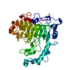
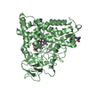
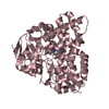
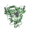
| Title | Structural insights into substrate specificity and the anti beta-elimination mechanism of pectate lyase | |||||||||
 Components Components | Pectate lyase | |||||||||
 Keywords Keywords | LYASE / Michaelis complex / hexasaccharide / pectate lyase / Calcium / Secreted | |||||||||
| Function / homology |  Function and homology information Function and homology informationpectate lyase / pectate lyase activity / pectin catabolic process / extracellular region / metal ion binding Similarity search - Function | |||||||||
| Biological species |  | |||||||||
| Method |  X-RAY DIFFRACTION / SYNCHROTRON / FOURIER SYNTHESIS / Resolution: 1.9 Å X-RAY DIFFRACTION / SYNCHROTRON / FOURIER SYNTHESIS / Resolution: 1.9 Å | |||||||||
 Authors Authors | Pickersgill, R.W. / To, T.T. | |||||||||
 Citation Citation | Journal: Biochemistry / Year: 2010 Title: Structural insights into substrate specificity and the anti beta-elimination mechanism of pectate lyase Authors: Seyedarabi, A. / To, T.T. / Ali, S. / Hussain, S. / Fries, M. / Madsen, R. / Clausen, M.H. / Teixteira, S. / Brocklehurst, K. / Pickersgill, R.W. | |||||||||
| History |
|
- Structure visualization
| Structure viewer | Molecule: MolmilJmol/JSmol |
|---|
- Downloads & links
-Download
| PDBx/mmCIF format | 3krg.cif.gz | 110.4 KB | Display | PDBx/mmCIF format |
|---|---|---|---|---|
| PDB format | pdb3krg.ent.gz | 80.2 KB | Display | PDB format |
| PDBx/mmJSON format | 3krg.json.gz | Tree view | PDBx/mmJSON format | |
| Others |  Other downloads Other downloads |
-Validation report
| Arichive directory | https://data.pdbj.org/pub/pdb/validation_reports/kr/3krgftp://data.pdbj.org/pub/pdb/validation_reports/kr/3krg | HTTPS FTP |
|---|
-Related structure data
| Related structure data |  2nzmC  2o04C  2o0vSC  2o17C  2o1dC C: citing same article ( S: Starting model for refinement |
|---|---|
| Similar structure data |


-Links
 PDBj
PDBj
- Assembly
| Deposited unit | 
| ||||||||
|---|---|---|---|---|---|---|---|---|---|
| 1 |
| ||||||||
| Unit cell |
|
-Components
| #1: Protein | Mass: 43300.059 Da / Num. of mol.: 1 / Mutation: D173A,N180A,K247A Source method: isolated from a genetically manipulated source Source: (gene. exp.) |
|---|---|
| #2: Polysaccharide | alpha-D-galactopyranuronic acid-(1-4)-alpha-D-galactopyranuronic acid-(1-4)-alpha-D- ...alpha-D-galactopyranuronic acid-(1-4)-alpha-D-galactopyranuronic acid-(1-4)-alpha-D-galactopyranuronic acid-(1-4)-alpha-D-galactopyranuronic acid-(1-4)-alpha-D-galactopyranuronic acid-(1-4)-alpha-D-galactopyranuronic acid Source method: isolated from a genetically manipulated source |
| #3: Chemical | ChemComp-CO /   Mass: 58.933 Da / Num. of mol.: 1 / Source method: obtained synthetically / Formula: Co Mass: 58.933 Da / Num. of mol.: 1 / Source method: obtained synthetically / Formula: Co |
| #4: Chemical | ChemComp-GOL /   Mass: 92.094 Da / Num. of mol.: 1 / Source method: obtained synthetically / Formula: C3H8O3 Mass: 92.094 Da / Num. of mol.: 1 / Source method: obtained synthetically / Formula: C3H8O3 |
| #5: Water | ChemComp-HOH /  Mass: 18.015 Da / Num. of mol.: 723 / Source method: isolated from a natural source / Formula: H2O Mass: 18.015 Da / Num. of mol.: 723 / Source method: isolated from a natural source / Formula: H2O |
-Experimental details
-Experiment
| Experiment | Method: X-RAY DIFFRACTION / Number of used crystals: 1 |
|---|
- Sample preparation
| Crystal | Density Matthews: 2.2 Å3/Da / Density % sol: 44.17 % |
|---|---|
| Crystal grow | Temperature: 293 K / Method: vapor diffusion, hanging drop / pH: 4.6 Details: 25% PEG 4000, 0.2M ammonium acetate, 0.1M sodium acetate, 50% PEG, pH 4.6, VAPOR DIFFUSION, HANGING DROP, temperature 293K |
-Data collection
| Diffraction | Mean temperature: 100 K |
|---|---|
| Diffraction source | Source: SYNCHROTRON / Site: SRS  / Beamline: PX10.1 / Wavelength: 0.97 Å / Beamline: PX10.1 / Wavelength: 0.97 Å |
| Detector | Type: MARMOSAIC 225 mm CCD / Detector: CCD / Date: Jan 31, 2007 / Details: mirrors |
| Radiation | Protocol: SINGLE WAVELENGTH / Monochromatic (M) / Laue (L): M / Scattering type: x-ray |
| Radiation wavelength | Wavelength: 0.97 Å / Relative weight: 1 |
| Reflection | Resolution: 1.9→69.01 Å / Num. obs: 28827 / % possible obs: 97.2 % / Observed criterion σ(F): 0 / Observed criterion σ(I): 0 / Redundancy: 3.7 % / Biso Wilson estimate: 8.608 Å2 / Rmerge(I) obs: 0.057 / Rsym value: 0.067 / Net I/σ(I): 23.8 |
| Reflection shell | Resolution: 1.9→2 Å / Redundancy: 3.7 % / Rmerge(I) obs: 0.067 / Mean I/σ(I) obs: 18.3 / Num. unique all: 4138 / Rsym value: 0.067 / % possible all: 96.1 |
- Processing
| Software |
| |||||||||||||||||||||||||||||||||||||||||||||||||||||||||||||||||
|---|---|---|---|---|---|---|---|---|---|---|---|---|---|---|---|---|---|---|---|---|---|---|---|---|---|---|---|---|---|---|---|---|---|---|---|---|---|---|---|---|---|---|---|---|---|---|---|---|---|---|---|---|---|---|---|---|---|---|---|---|---|---|---|---|---|---|
| Refinement | Method to determine structure: FOURIER SYNTHESIS Starting model: PDB entry 2O0V Resolution: 1.9→29.18 Å / Cor.coef. Fo:Fc: 0.952 / Cor.coef. Fo:Fc free: 0.911 / Occupancy max: 1 / Occupancy min: 1 / SU B: 2.464 / SU ML: 0.076 / Isotropic thermal model: Isotropic / Cross valid method: THROUGHOUT / ESU R: 0.147 / ESU R Free: 0.144 / Stereochemistry target values: MAXIMUM LIKELIHOOD Details: HYDROGENS HAVE BEEN ADDED IN THE RIDING POSITIONS U VALUES: REFINED INDIVIDUALLY
| |||||||||||||||||||||||||||||||||||||||||||||||||||||||||||||||||
| Solvent computation | Ion probe radii: 0.8 Å / Shrinkage radii: 0.8 Å / VDW probe radii: 1.4 Å / Solvent model: MASK | |||||||||||||||||||||||||||||||||||||||||||||||||||||||||||||||||
| Displacement parameters | Biso max: 66.69 Å2 / Biso mean: 8.969 Å2 / Biso min: 2 Å2
| |||||||||||||||||||||||||||||||||||||||||||||||||||||||||||||||||
| Refinement step | Cycle: LAST / Resolution: 1.9→29.18 Å
| |||||||||||||||||||||||||||||||||||||||||||||||||||||||||||||||||
| Refine LS restraints |
| |||||||||||||||||||||||||||||||||||||||||||||||||||||||||||||||||
| LS refinement shell | Resolution: 0.129→1.9 Å / Total num. of bins used: 20
|
