 Movie
Movie Controller


+ Open data

- Basic information
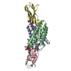
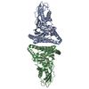
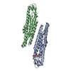
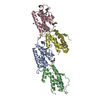
| Entry | Database: PDB / ID: 3kph | ||||||
|---|---|---|---|---|---|---|---|
| Title | Crystal structure of Mycoplasma arthritidis-derived mitogen | ||||||
 Components Components | Superantigen | ||||||
 Keywords Keywords | IMMUNE SYSTEM / superantigen / MAM / 3D-domain swap | ||||||
| Function / homology |  Function and homology information Function and homology informationmam-mhc complex, Chain D, Domain 2 / Hla class ii histocompatibility antigen, dr alpha chain. Chain D, domain 1 / Mycoplasma arthritidis-derived mitogen / Superantigen MAM / Mycoplasma arthritidis-derived mitogen, C-terminal / Mycoplasma arthritidis-derived mitogen / Four Helix Bundle (Hemerythrin (Met), subunit A) / Arc Repressor Mutant, subunit A / Up-down Bundle / Orthogonal Bundle / Mainly Alpha Similarity search - Domain/homology | ||||||
| Biological species |  Mycoplasma arthritidis (bacteria) Mycoplasma arthritidis (bacteria) | ||||||
| Method |  X-RAY DIFFRACTION / SYNCHROTRON / MOLECULAR REPLACEMENT / Resolution: 2.8 Å X-RAY DIFFRACTION / SYNCHROTRON / MOLECULAR REPLACEMENT / Resolution: 2.8 Å | ||||||
 Authors Authors | Liu, L.H. / Li, H.M. | ||||||
 Citation Citation | Journal: J.Mol.Biol. / Year: 2010 Title: Crystal Structure of the Mycoplasma arthritidis-Derived Mitogen in Apo Form Reveals a 3D Domain-Swapped Dimer. Authors: Liu, L. / Li, Z. / Guo, Y. / Vanvranken, S.J. / Mourad, W. / Li, H. | ||||||
| History |
|
- Structure visualization
| Structure viewer | Molecule: MolmilJmol/JSmol |
|---|
- Downloads & links
-Download
| PDBx/mmCIF format | 3kph.cif.gz | 93.6 KB | Display | PDBx/mmCIF format |
|---|---|---|---|---|
| PDB format | pdb3kph.ent.gz | 72.7 KB | Display | PDB format |
| PDBx/mmJSON format | 3kph.json.gz | Tree view | PDBx/mmJSON format | |
| Others |  Other downloads Other downloads |
-Validation report
| Arichive directory | https://data.pdbj.org/pub/pdb/validation_reports/kp/3kphftp://data.pdbj.org/pub/pdb/validation_reports/kp/3kph | HTTPS FTP |
|---|
-Related structure data
| Related structure data |  1r5iS S: Starting model for refinement |
|---|---|
| Similar structure data |


-Links
 PDBj
PDBj- Assembly
| Deposited unit | 
| ||||||||
|---|---|---|---|---|---|---|---|---|---|
| 1 |
| ||||||||
| Unit cell |
| ||||||||
| Components on special symmetry positions |
|
-Components
| #1: Protein | Mass: 25570.594 Da / Num. of mol.: 2 / Fragment: UNP residues 26-239 / Mutation: K201A Source method: isolated from a genetically manipulated source Source: (gene. exp.) Mycoplasma arthritidis (bacteria) / Plasmid: pGEX-6P-1 / Production host: #2: Chemical |   Mass: 94.971 Da / Num. of mol.: 3 / Source method: obtained synthetically / Formula: PO4 Mass: 94.971 Da / Num. of mol.: 3 / Source method: obtained synthetically / Formula: PO4#3: Water | ChemComp-HOH / |  Mass: 18.015 Da / Num. of mol.: 62 / Source method: isolated from a natural source / Formula: H2O Mass: 18.015 Da / Num. of mol.: 62 / Source method: isolated from a natural source / Formula: H2O |
|---|
-Experimental details
-Experiment
| Experiment | Method: X-RAY DIFFRACTION / Number of used crystals: 1 |
|---|
- Sample preparation
| Crystal | Density Matthews: 4.9 Å3/Da / Density % sol: 74.9 % |
|---|---|
| Crystal grow | Temperature: 293 K / Method: vapor diffusion / pH: 6.2 Details: 13-15% PEG 3350, 0.2 M NaCl, 5% Ethylene glycol, 5% Glycerol, 0.1 M Potassium/sodium phosphate pH 6.2, VAPOR DIFFUSION, temperature 293K |
-Data collection
| Diffraction | Mean temperature: 100 K |
|---|---|
| Diffraction source | Source: SYNCHROTRON / Site: NSLS  / Beamline: X4A / Wavelength: 1.2757 Å / Beamline: X4A / Wavelength: 1.2757 Å |
| Detector | Type: ADSC QUANTUM 4 / Detector: CCD / Date: Sep 10, 2004 / Details: Mirrors |
| Radiation | Protocol: SINGLE WAVELENGTH / Monochromatic (M) / Laue (L): M / Scattering type: x-ray |
| Radiation wavelength | Wavelength: 1.2757 Å / Relative weight: 1 |
| Reflection | Resolution: 2.8→50 Å / Num. all: 24867 / Num. obs: 24867 / % possible obs: 95.9 % / Observed criterion σ(I): 0 / Redundancy: 6.93 % / Rmerge(I) obs: 0.117 / Rsym value: 0.117 / Net I/σ(I): 9.6 |
| Reflection shell | Resolution: 2.8→2.9 Å / Redundancy: 5.4 % / Rmerge(I) obs: 0.692 / Mean I/σ(I) obs: 2.7 / Rsym value: 0.692 / % possible all: 96.8 |
- Processing
| Software |
| ||||||||||||||||||||||||||||||||||||
|---|---|---|---|---|---|---|---|---|---|---|---|---|---|---|---|---|---|---|---|---|---|---|---|---|---|---|---|---|---|---|---|---|---|---|---|---|---|
| Refinement | Method to determine structure: MOLECULAR REPLACEMENT Starting model: PDB entry 1R5I Resolution: 2.8→50 Å / Rfactor Rfree error: 0.008 / Occupancy max: 1 / Occupancy min: 0.5 / Data cutoff high absF: 2330671 / Data cutoff low absF: 0 / Isotropic thermal model: RESTRAINED / Cross valid method: THROUGHOUT / σ(F): 0 / σ(I): 0 / Stereochemistry target values: Engh & Huber
| ||||||||||||||||||||||||||||||||||||
| Solvent computation | Solvent model: FLAT MODEL / Bsol: 50.483 Å2 / ksol: 0.353 e/Å3 | ||||||||||||||||||||||||||||||||||||
| Displacement parameters | Biso max: 133.23 Å2 / Biso mean: 69.238 Å2 / Biso min: 19.64 Å2
| ||||||||||||||||||||||||||||||||||||
| Refine analyze |
| ||||||||||||||||||||||||||||||||||||
| Refinement step | Cycle: LAST / Resolution: 2.8→50 Å
| ||||||||||||||||||||||||||||||||||||
| Refine LS restraints |
| ||||||||||||||||||||||||||||||||||||
| LS refinement shell | Resolution: 2.8→2.98 Å / Rfactor Rfree error: 0.028 / Total num. of bins used: 6
| ||||||||||||||||||||||||||||||||||||
| Xplor file |
|
