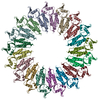
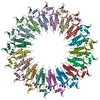
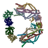
A: Coat protein B: Coat protein C: Coat protein D: Coat protein E: Coat protein F: Coat protein G: Coat protein H: Coat protein I: Coat protein J: Coat protein K: Coat protein L: Coat protein M: Coat protein N: Coat protein O: Coat protein P: Coat protein Q: Coat protein
A: Coat protein B: Coat protein C: Coat protein D: Coat protein E: Coat protein F: Coat protein G: Coat protein H: Coat protein I: Coat protein J: Coat protein K: Coat protein L: Coat protein M: Coat protein N: Coat protein O: Coat protein P: Coat protein Q: Coat protein
A: Coat protein B: Coat protein C: Coat protein D: Coat protein E: Coat protein F: Coat protein G: Coat protein H: Coat protein I: Coat protein J: Coat protein K: Coat protein L: Coat protein M: Coat protein N: Coat protein O: Coat protein P: Coat protein Q: Coat protein
 Movie
Movie Controller


 Open data
 Basic information
 Components
 Keywords
 Function and homology information
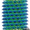 Tobacco mosaic virus
 X-RAY DIFFRACTION /
  Authors
 Citation
 Structure visualization
 Downloads & links
 Other downloads


 PDBj
PDBj
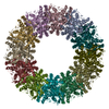
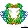
 Assembly