 Movie
Movie Controller


+ Open data

- Basic information
| Entry | Database: PDB / ID: 3kin | ||||||
|---|---|---|---|---|---|---|---|
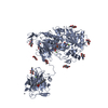
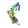
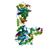
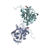
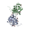
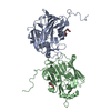
| Title | KINESIN (DIMERIC) FROM RATTUS NORVEGICUS | ||||||
 Components Components | (KINESIN HEAVY CHAIN) x 2 | ||||||
 Keywords Keywords | MOTOR PROTEIN / CYTOSKELETON | ||||||
| Function / homology |  Function and homology information Function and homology informationresponse to rotenone / RHO GTPases activate KTN1 / Kinesins / distal axon / anterograde dendritic transport of messenger ribonucleoprotein complex / COPI-dependent Golgi-to-ER retrograde traffic / plus-end-directed kinesin ATPase / anterograde dendritic transport of neurotransmitter receptor complex / central region of growth cone / anterograde axonal protein transport ...response to rotenone / RHO GTPases activate KTN1 / Kinesins / distal axon / anterograde dendritic transport of messenger ribonucleoprotein complex / COPI-dependent Golgi-to-ER retrograde traffic / plus-end-directed kinesin ATPase / anterograde dendritic transport of neurotransmitter receptor complex / central region of growth cone / anterograde axonal protein transport / MHC class II antigen presentation / retrograde neuronal dense core vesicle transport / apolipoprotein receptor binding / ciliary rootlet / intracellular mRNA localization / thalamus development / plus-end-directed microtubule motor activity / apical dendrite / motor neuron axon guidance / microtubule motor activity / kinesin complex / synaptic vesicle transport / cellular response to ethanol / kinesin binding / mRNA transport / postsynaptic cytosol / axonal growth cone / neuron development / microtubule-based process / vesicle-mediated transport / axon cytoplasm / dendrite cytoplasm / axon guidance / hippocampus development / P-body / cellular response to nerve growth factor stimulus / cerebral cortex development / Hydrolases; Acting on acid anhydrides; Acting on acid anhydrides to facilitate cellular and subcellular movement / GABA-ergic synapse / scaffold protein binding / microtubule binding / microtubule / perikaryon / neuron projection / axon / neuronal cell body / dendrite / protein-containing complex binding / perinuclear region of cytoplasm / ATP hydrolysis activity / ATP binding / cytoplasm Similarity search - Function | ||||||
| Biological species |  | ||||||
| Method |  X-RAY DIFFRACTION / SYNCHROTRON / MIRAS / Resolution: 3.1 Å X-RAY DIFFRACTION / SYNCHROTRON / MIRAS / Resolution: 3.1 Å | ||||||
 Authors Authors | Kozielski, F. / Sack, S. / Marx, A. / Thormahlen, M. / Schonbrunn, E. / Biou, V. / Thompson, A. / Mandelkow, E.-M. / Mandelkow, E. | ||||||
 Citation Citation | Journal: Cell(Cambridge,Mass.) / Year: 1997 Title: The crystal structure of dimeric kinesin and implications for microtubule-dependent motility. Authors: Kozielski, F. / Sack, S. / Marx, A. / Thormahlen, M. / Schonbrunn, E. / Biou, V. / Thompson, A. / Mandelkow, E.M. / Mandelkow, E. #1: Journal: J.Struct.Biol. / Year: 1997Title: Crystallization and Preliminary X-Ray Analysis of the Single-Headed and Double-Headed Motor Protein Kinesin Authors: Kozielski, F. / Schonbrunn, E. / Sack, S. / Muller, J. / Brady, S.T. / Mandelkow, E. #2: Journal: Biochemistry / Year: 1997Title: X-Ray Structure of Motor and Neck Domains from Rat Brain Kinesin Authors: Sack, S. / Muller, J. / Marx, A. / Thormahlen, M. / Mandelkow, E.M. / Brady, S.T. / Mandelkow, E. | ||||||
| History |
|
- Structure visualization
| Structure viewer | Molecule: MolmilJmol/JSmol |
|---|
- Downloads & links
-Download
| PDBx/mmCIF format | 3kin.cif.gz | 137.1 KB | Display | PDBx/mmCIF format |
|---|---|---|---|---|
| PDB format | pdb3kin.ent.gz | 105.5 KB | Display | PDB format |
| PDBx/mmJSON format | 3kin.json.gz | Tree view | PDBx/mmJSON format | |
| Others |  Other downloads Other downloads |
-Validation report
| Arichive directory | https://data.pdbj.org/pub/pdb/validation_reports/ki/3kinftp://data.pdbj.org/pub/pdb/validation_reports/ki/3kin | HTTPS FTP |
|---|
-Related structure data
| Similar structure data |
|---|


-Links
 PDBj
PDBj


- Assembly
| Deposited unit | 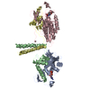
| ||||||||
|---|---|---|---|---|---|---|---|---|---|
| 1 |
| ||||||||
| 2 | 
| ||||||||
| Unit cell |
|
-Components
| #1: Protein | Mass: 27072.869 Da / Num. of mol.: 2 / Fragment: MOTOR DOMAIN Source method: isolated from a genetically manipulated source Source: (gene. exp.)  #2: Protein | Mass: 13248.242 Da / Num. of mol.: 2 / Fragment: MOTOR DOMAIN Source method: isolated from a genetically manipulated source Source: (gene. exp.) #3: Chemical |   Mass: 427.201 Da / Num. of mol.: 2 / Source method: obtained synthetically / Formula: C10H15N5O10P2 / Comment: ADP, energy-carrying molecule*YM Mass: 427.201 Da / Num. of mol.: 2 / Source method: obtained synthetically / Formula: C10H15N5O10P2 / Comment: ADP, energy-carrying molecule*YM#4: Water | ChemComp-HOH / |  Mass: 18.015 Da / Num. of mol.: 60 / Source method: isolated from a natural source / Formula: H2O Mass: 18.015 Da / Num. of mol.: 60 / Source method: isolated from a natural source / Formula: H2OHas protein modification | N | |
|---|
-Experimental details
-Experiment
| Experiment | Method: X-RAY DIFFRACTION / Number of used crystals: 1 |
|---|
- Sample preparation
| Crystal | Density Matthews: 2.8 Å3/Da / Density % sol: 55 % | ||||||||||||||||||||||||||||||||||||||||||||||||
|---|---|---|---|---|---|---|---|---|---|---|---|---|---|---|---|---|---|---|---|---|---|---|---|---|---|---|---|---|---|---|---|---|---|---|---|---|---|---|---|---|---|---|---|---|---|---|---|---|---|
| Crystal grow | Method: vapor diffusion, hanging drop / pH: 7.5 Details: HANGING DROP METHOD WAS USED. PROTEIN WAS CRYSTALLIZED FROM 20MM PIPES, PH 7.5, 200MM NACL, 2MM DTT, 2 MM NA-EGTA, 0.8 M AS. THE RESERVOIR CONTAINED 1.6 M AS IN THE SAME BUFFER., vapor diffusion - hanging drop | ||||||||||||||||||||||||||||||||||||||||||||||||
| Crystal | *PLUS | ||||||||||||||||||||||||||||||||||||||||||||||||
| Crystal grow | *PLUS Temperature: 19 ℃ / Method: vapor diffusion, hanging drop | ||||||||||||||||||||||||||||||||||||||||||||||||
| Components of the solutions | *PLUS
|
-Data collection
| Diffraction | Mean temperature: 90 K |
|---|---|
| Diffraction source | Source: SYNCHROTRON / Site: ESRF  / Beamline: BM14 / Wavelength: 0.92 / Beamline: BM14 / Wavelength: 0.92 |
| Detector | Type: MARRESEARCH / Detector: IMAGE PLATE / Date: Jul 1, 1996 / Details: MIRRORS |
| Radiation | Monochromator: SI(111) / Monochromatic (M) / Laue (L): M / Scattering type: x-ray |
| Radiation wavelength | Wavelength: 0.92 Å / Relative weight: 1 |
| Reflection | Resolution: 3→30 Å / Num. obs: 19516 / % possible obs: 100 % / Rsym value: 0.064 / Net I/σ(I): 10.7 |
| Reflection | *PLUS Num. measured all: 113071 / Rmerge(I) obs: 0.064 |
- Processing
| Software |
| |||||||||||||||
|---|---|---|---|---|---|---|---|---|---|---|---|---|---|---|---|---|
| Refinement | Method to determine structure: MIRAS / Resolution: 3.1→6 Å / Cross valid method: FREE R
| |||||||||||||||
| Refinement step | Cycle: LAST / Resolution: 3.1→6 Å
|
