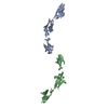
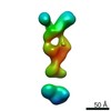
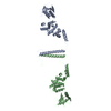
- PDB-3k8p: Structural basis for vesicle tethering by the Dsl1 complex -
+
Open data
ID or keywords:
Loading...
-
Basic information
Entry
Database: PDB / ID: 3k8p
Title
Structural basis for vesicle tethering by the Dsl1 complex
Components
Dsl1
Protein transport protein SEC39
Keywords
TRANSPORT PROTEIN/TRANSPORT PROTEIN / Intracellular trafficking / Dsl1 complex / multisubunit tethering complex / SNARE proteins / Endoplasmic reticulum / ER-Golgi transport / Membrane / Protein transport / Transport / TRANSPORT PROTEIN-TRANSPORT PROTEIN complex
Function / homology
Function and homology information
ER-dependent peroxisome organization / Dsl1/NZR complex / RZZ complex / retrograde vesicle-mediated transport, Golgi to endoplasmic reticulum / mitotic spindle assembly checkpoint signaling / endoplasmic reticulum to Golgi vesicle-mediated transport / vesicle-mediated transport / nuclear envelope / protein transport / endoplasmic reticulum membrane ...ER-dependent peroxisome organization / Dsl1/NZR complex / RZZ complex / retrograde vesicle-mediated transport, Golgi to endoplasmic reticulum / mitotic spindle assembly checkpoint signaling / endoplasmic reticulum to Golgi vesicle-mediated transport / vesicle-mediated transport / nuclear envelope / protein transport / endoplasmic reticulum membrane / endoplasmic reticulum / cytoplasm Similarity search - Function
Methane Monooxygenase Hydroxylase; Chain G, domain 1 - #1440 / Tetracycline Repressor; domain 2 - #150 / Sec39 domain / Secretory pathway protein Sec39 / Retrograde transport protein Dsl1 N-terminal domain / Retrograde transport protein Dsl1, C-terminal domain / Dsl1, N-terminal domain superfamily / Retrograde transport protein Dsl1 N terminal / Retrograde transport protein Dsl1 C terminal / Zw10/DSL1, C-terminal ...Methane Monooxygenase Hydroxylase; Chain G, domain 1 - #1440 / Tetracycline Repressor; domain 2 - #150 / Sec39 domain / Secretory pathway protein Sec39 / Retrograde transport protein Dsl1 N-terminal domain / Retrograde transport protein Dsl1, C-terminal domain / Dsl1, N-terminal domain superfamily / Retrograde transport protein Dsl1 N terminal / Retrograde transport protein Dsl1 C terminal / Zw10/DSL1, C-terminal / Tetracycline Repressor; domain 2 / Methane Monooxygenase Hydroxylase; Chain G, domain 1 / Up-down Bundle / Orthogonal Bundle / Mainly Alpha Similarity search - Domain/homology
In the structure databanks used in Yorodumi, some data are registered as the other names, "COVID-19 virus" and "2019-nCoV". Here are the details of the virus and the list of structure data.
Jan 31, 2019. EMDB accession codes are about to change! (news from PDBe EMDB page)
EMDB accession codes are about to change! (news from PDBe EMDB page)
The allocation of 4 digits for EMDB accession codes will soon come to an end. Whilst these codes will remain in use, new EMDB accession codes will include an additional digit and will expand incrementally as the available range of codes is exhausted. The current 4-digit format prefixed with “EMD-” (i.e. EMD-XXXX) will advance to a 5-digit format (i.e. EMD-XXXXX), and so on. It is currently estimated that the 4-digit codes will be depleted around Spring 2019, at which point the 5-digit format will come into force.
The EM Navigator/Yorodumi systems omit the EMD- prefix.
Related info.:Q: What is EMD? / ID/Accession-code notation in Yorodumi/EM Navigator
Yorodumi is a browser for structure data from EMDB, PDB, SASBDB, etc.
This page is also the successor to EM Navigator detail page, and also detail information page/front-end page for Omokage search.
The word "yorodu" (or yorozu) is an old Japanese word meaning "ten thousand". "mi" (miru) is to see.
Related info.:EMDB / PDB / SASBDB / Comparison of 3 databanks / Yorodumi Search / Aug 31, 2016. New EM Navigator & Yorodumi / Yorodumi Papers / Jmol/JSmol / Function and homology information / Changes in new EM Navigator and Yorodumi
 Movie
Movie Controller


 Open data
 Basic information
 Components
 Keywords
 Function and homology information
 Kluyveromyces lactis (yeast)
 X-RAY DIFFRACTION /
  Authors
 Citation
 Structure visualization
 Downloads & links
 Other downloads
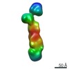


 PDBj
PDBj
 Assembly

 Mass: 18.015 Da / Num. of mol.: 3 / Source method: isolated from a natural source / Formula: H2O
 Sample preparation
 / Beamline: X29A / Wavelength: 0.9793, 0.9795, 0.9641
 Processing