 Movie
Movie Controller

[English] 日本語
 Yorodumi
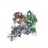
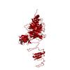
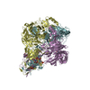
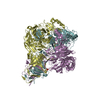
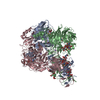
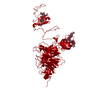
- PDB-3ju4: Crystal Structure Analysis of EndosialidaseNF at 0.98 A Resolution -
+ Open data

- Basic information
| Entry | Database: PDB / ID: 3ju4 | ||||||
|---|---|---|---|---|---|---|---|
| Title | Crystal Structure Analysis of EndosialidaseNF at 0.98 A Resolution | ||||||
 Components Components | Endo-N-acetylneuraminidase | ||||||
 Keywords Keywords | HYDROLASE / endoNF / polySia / high-resolution / 1A / Glycosidase | ||||||
| Function / homology |  Function and homology information Function and homology informationendo-alpha-sialidase / endo-alpha-(2,8)-sialidase activity / symbiont entry into host cell via disruption of host cell glycocalyx / virus tail, fiber / symbiont entry into host cell via disruption of host cell envelope / symbiont entry into host / adhesion receptor-mediated virion attachment to host cell / virion attachment to host cell / identical protein binding Similarity search - Function | ||||||
| Biological species |  Enterobacteria phage K1F (virus) Enterobacteria phage K1F (virus) | ||||||
| Method |  X-RAY DIFFRACTION / SYNCHROTRON / MOLECULAR REPLACEMENT / Resolution: 0.98 Å X-RAY DIFFRACTION / SYNCHROTRON / MOLECULAR REPLACEMENT / Resolution: 0.98 Å | ||||||
 Authors Authors | Schulz, E.C. / Neuman, P. / Gerardy-Schahn, R. / Sheldrick, G.M. / Ficner, R. | ||||||
 Citation Citation | Journal: Acta Crystallogr.,Sect.D / Year: 2010 Title: Structure analysis of endosialidase NF at 0.98 A resolution. Authors: Schulz, E.C. / Neumann, P. / Gerardy-Schahn, R. / Sheldrick, G.M. / Ficner, R. | ||||||
| History |
|
- Structure visualization
| Structure viewer | Molecule: MolmilJmol/JSmol |
|---|
- Downloads & links
-Download
| PDBx/mmCIF format | 3ju4.cif.gz | 356.3 KB | Display | PDBx/mmCIF format |
|---|---|---|---|---|
| PDB format | pdb3ju4.ent.gz | 284 KB | Display | PDB format |
| PDBx/mmJSON format | 3ju4.json.gz | Tree view | PDBx/mmJSON format | |
| Others |  Other downloads Other downloads |
-Validation report
| Summary document | 3ju4_validation.pdf.gz | 779.3 KB | Display | wwPDB validaton report |
|---|---|---|---|---|
| Full document | 3ju4_full_validation.pdf.gz | 787.8 KB | Display | |
| Data in XML | 3ju4_validation.xml.gz | 43.1 KB | Display | |
| Data in CIF | 3ju4_validation.cif.gz | 72.9 KB | Display | |
| Arichive directory | https://data.pdbj.org/pub/pdb/validation_reports/ju/3ju4ftp://data.pdbj.org/pub/pdb/validation_reports/ju/3ju4 | HTTPS FTP |
-Related structure data
| Related structure data | 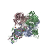 1v0eS S: Starting model for refinement |
|---|---|
| Similar structure data |


-Links
 PDBj
PDBj


- Assembly
| Deposited unit | 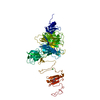
| ||||||||||||
|---|---|---|---|---|---|---|---|---|---|---|---|---|---|
| 1 | 
| ||||||||||||
| Unit cell |
| ||||||||||||
| Components on special symmetry positions |
|
-Components
| #1: Protein | Mass: 74542.461 Da / Num. of mol.: 1 / Fragment: UNP RESIDUES 246-910 Source method: isolated from a genetically manipulated source Source: (gene. exp.) Enterobacteria phage K1F (virus) / Strain: K1 / Plasmid: pet22b+ / Production host:  | ||||||
|---|---|---|---|---|---|---|---|
| #2: Sugar | ChemComp-SLB / 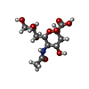  Type: D-saccharide, beta linking / Mass: 309.270 Da / Num. of mol.: 1 Type: D-saccharide, beta linking / Mass: 309.270 Da / Num. of mol.: 1Source method: isolated from a genetically manipulated source Formula: C11H19NO9 | ||||||
| #3: Chemical | ChemComp-CL /   Mass: 35.453 Da / Num. of mol.: 5 / Source method: obtained synthetically / Formula: Cl Mass: 35.453 Da / Num. of mol.: 5 / Source method: obtained synthetically / Formula: Cl#4: Chemical | ChemComp-NA / |   Mass: 22.990 Da / Num. of mol.: 1 / Source method: obtained synthetically / Formula: Na Mass: 22.990 Da / Num. of mol.: 1 / Source method: obtained synthetically / Formula: Na#5: Water | ChemComp-HOH / |  Mass: 18.015 Da / Num. of mol.: 1616 / Source method: isolated from a natural source / Formula: H2O Mass: 18.015 Da / Num. of mol.: 1616 / Source method: isolated from a natural source / Formula: H2OSequence details | THE DEPOSITORS | |
-Experimental details
-Experiment
| Experiment | Method: X-RAY DIFFRACTION / Number of used crystals: 1 |
|---|
- Sample preparation
| Crystal | Density Matthews: 3.21 Å3/Da / Density % sol: 61.73 % |
|---|---|
| Crystal grow | Temperature: 293 K / Method: vapor diffusion / pH: 7.2 Details: 16% (w/v) PEG 8000, 0.1M Tris/HCl pH 7.2, 3% Isopropanol, VAPOR DIFFUSION, temperature 293K |
-Data collection
| Diffraction | Mean temperature: 100 K |
|---|---|
| Diffraction source | Source: SYNCHROTRON / Site: ESRF  / Beamline: ID14-1 / Wavelength: 0.934 Å / Beamline: ID14-1 / Wavelength: 0.934 Å |
| Detector | Type: ADSC QUANTUM 210 / Detector: CCD / Date: Sep 20, 2008 |
| Radiation | Protocol: SINGLE WAVELENGTH / Monochromatic (M) / Laue (L): M / Scattering type: x-ray |
| Radiation wavelength | Wavelength: 0.934 Å / Relative weight: 1 |
| Reflection | Resolution: 0.98→20 Å / Num. obs: 516832 / % possible obs: 96.9 % / Redundancy: 3.3 % / Rsym value: 0.073 / Net I/σ(I): 12.41 |
| Reflection shell | Resolution: 0.98→1.08 Å / Redundancy: 3.2 % / Mean I/σ(I) obs: 3.42 / Num. unique all: 429843 / Rsym value: 0.415 / % possible all: 99.6 |
- Processing
| Software |
| ||||||||||||||||||||
|---|---|---|---|---|---|---|---|---|---|---|---|---|---|---|---|---|---|---|---|---|---|
| Refinement | Method to determine structure: MOLECULAR REPLACEMENT Starting model: 1V0E Resolution: 0.98→20 Å / Cross valid method: THROUGHOUT / σ(F): 0 / Stereochemistry target values: Engh & Huber
| ||||||||||||||||||||
| Refine analyze | Num. disordered residues: 43 | ||||||||||||||||||||
| Refinement step | Cycle: LAST / Resolution: 0.98→20 Å
|
