 Movie
Movie Controller

[English] 日本語
 Yorodumi
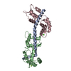
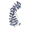
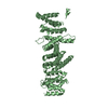
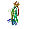
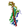
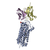
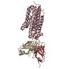
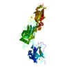
- PDB-3jtd: Calcium-free Scallop Myosin Regulatory Domain with ELC-D19A Point... -
+ Open data

- Basic information
| Entry | Database: PDB / ID: 3jtd | ||||||
|---|---|---|---|---|---|---|---|
| Title | Calcium-free Scallop Myosin Regulatory Domain with ELC-D19A Point Mutation | ||||||
 Components Components |
| ||||||
 Keywords Keywords | CONTRACTILE PROTEIN / Regulated myosins / smooth and molluscan muscle / X-ray crystallographic structure / scallop regulatory domain/lever arm / off-state / Actin-binding / ATP-binding / Calmodulin-binding / Coiled coil / Cytoplasm / Motor protein / Muscle protein / Myosin / Nucleotide-binding / Thick filament / Calcium | ||||||
| Function / homology |  Function and homology information Function and homology informationmyosin filament organization / myofibril assembly / muscle myosin complex / myosin filament / locomotion / myosin complex / myosin II complex / structural constituent of muscle / microfilament motor activity / myofibril ...myosin filament organization / myofibril assembly / muscle myosin complex / myosin filament / locomotion / myosin complex / myosin II complex / structural constituent of muscle / microfilament motor activity / myofibril / muscle contraction / actin filament binding / calmodulin binding / calcium ion binding / ATP binding Similarity search - Function | ||||||
| Biological species |  Argopecten irradians (bay scallop) Argopecten irradians (bay scallop) | ||||||
| Method |  X-RAY DIFFRACTION / SYNCHROTRON / MOLECULAR REPLACEMENT / molecular replacement / Resolution: 2.57 Å X-RAY DIFFRACTION / SYNCHROTRON / MOLECULAR REPLACEMENT / molecular replacement / Resolution: 2.57 Å | ||||||
 Authors Authors | Himmel, D.M. / Mui, S. / O'Neall-Hennessey, E. / Szent-Gyorgyi, A. / Cohen, C. | ||||||
 Citation Citation | Journal: J.Mol.Biol. / Year: 2009 Title: The on-off switch in regulated myosins: different triggers but related mechanisms. Authors: Himmel, D.M. / Mui, S. / O'Neall-Hennessey, E. / Szent-Gyorgyi, A.G. / Cohen, C. #1: Journal: Structure / Year: 1996Title: Structure of the Regulatory Domain of Scallop Myosin at 2 Resolution:Implications for Regulation. Authors: Houdusse, A. / Cohen, C. #2: Journal: Nature / Year: 1994Title: Structure of the Regulatory Domain of Scallop Myosin at 2.8 Resolution. Authors: Xie, X. / Harrison, D.H. / Schlichting, I. / Sweet, R.M. / Kalabokis, V.N. / Szent-Gy rgyi, A. / Cohen, C. | ||||||
| History |
|
- Structure visualization
| Structure viewer | Molecule: MolmilJmol/JSmol |
|---|
- Downloads & links
-Download
| PDBx/mmCIF format | 3jtd.cif.gz | 90.2 KB | Display | PDBx/mmCIF format |
|---|---|---|---|---|
| PDB format | pdb3jtd.ent.gz | 66.7 KB | Display | PDB format |
| PDBx/mmJSON format | 3jtd.json.gz | Tree view | PDBx/mmJSON format | |
| Others |  Other downloads Other downloads |
-Validation report
| Arichive directory | https://data.pdbj.org/pub/pdb/validation_reports/jt/3jtdftp://data.pdbj.org/pub/pdb/validation_reports/jt/3jtd | HTTPS FTP |
|---|
-Related structure data
| Related structure data |  3jvtC 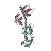 1wdcS S: Starting model for refinement C: citing same article ( |
|---|---|
| Similar structure data |


-Links
 PDBj
PDBj


- Assembly
| Deposited unit | 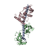
| ||||||||
|---|---|---|---|---|---|---|---|---|---|
| 1 |
| ||||||||
| Unit cell |
|
-Components
| #1: Protein | Mass: 8128.819 Da / Num. of mol.: 1 / Source method: isolated from a natural source / Source: (natural) Argopecten irradians (bay scallop) / References: UniProt: P24733 |
|---|---|
| #2: Protein | Mass: 17560.855 Da / Num. of mol.: 1 / Source method: isolated from a natural source / Source: (natural) Argopecten irradians (bay scallop) / References: UniProt: P13543 |
| #3: Protein | Mass: 17591.625 Da / Num. of mol.: 1 / Mutation: D19A Source method: isolated from a genetically manipulated source Source: (gene. exp.) Argopecten irradians (bay scallop) / Plasmid: pMW172 / Production host:  |
| #4: Chemical | ChemComp-MG /   Mass: 24.305 Da / Num. of mol.: 1 / Source method: obtained synthetically / Formula: Mg Mass: 24.305 Da / Num. of mol.: 1 / Source method: obtained synthetically / Formula: Mg |
| #5: Water | ChemComp-HOH /  Mass: 18.015 Da / Num. of mol.: 58 / Source method: isolated from a natural source / Formula: H2O Mass: 18.015 Da / Num. of mol.: 58 / Source method: isolated from a natural source / Formula: H2O |
-Experimental details
-Experiment
| Experiment | Method: X-RAY DIFFRACTION / Number of used crystals: 1 |
|---|
- Sample preparation
| Crystal | Density Matthews: 2.7 Å3/Da / Density % sol: 54.42 % |
|---|---|
| Crystal grow | Temperature: 277 K / Method: vapor diffusion, hanging drop / pH: 8.3 Details: 100 mM bicine pH 8.3, 45 mM ammonium sulfate, 5 mM magnesium chloride, 2.5 mM EGTA, 3.0 mM sodium azide, 15% (wt/vol) PEG 4000, 4% (vol/vol) PEG 200, 6% (wt/vol) 2,3-butanediol, Hanging drop ...Details: 100 mM bicine pH 8.3, 45 mM ammonium sulfate, 5 mM magnesium chloride, 2.5 mM EGTA, 3.0 mM sodium azide, 15% (wt/vol) PEG 4000, 4% (vol/vol) PEG 200, 6% (wt/vol) 2,3-butanediol, Hanging drop vapor diffusion, temperature 277K, VAPOR DIFFUSION, HANGING DROP |
-Data collection
| Diffraction | Mean temperature: 100 K |
|---|---|
| Diffraction source | Source: SYNCHROTRON / Site: CHESS  / Beamline: A1 / Wavelength: 0.908 Å / Beamline: A1 / Wavelength: 0.908 Å |
| Detector | Type: PRINCETON 2K / Detector: CCD / Date: Dec 25, 1997 |
| Radiation | Protocol: SINGLE WAVELENGTH / Scattering type: x-ray |
| Radiation wavelength | Wavelength: 0.908 Å / Relative weight: 1 |
| Reflection | Resolution: 2.57→37 Å / Num. all: 15190 / Num. obs: 13187 / % possible obs: 90.5 % / Observed criterion σ(F): 1 / Observed criterion σ(I): 1 / Redundancy: 2.45 % / Biso Wilson estimate: 42 Å2 / Rsym value: 0.064 / Net I/σ(I): 12.298 |
-Phasing
| Phasing | Method: molecular replacement |
|---|
- Processing
| Software |
| ||||||||||||||||||||||||||||||||||||
|---|---|---|---|---|---|---|---|---|---|---|---|---|---|---|---|---|---|---|---|---|---|---|---|---|---|---|---|---|---|---|---|---|---|---|---|---|---|
| Refinement | Method to determine structure: MOLECULAR REPLACEMENT Starting model: PDB ENTRY 1WDC Resolution: 2.57→34.89 Å / Rfactor Rfree error: 0.01 / Occupancy max: 1 / Occupancy min: 0 / FOM work R set: 0.819 / Data cutoff high absF: 186917 / Data cutoff low absF: 0 / Isotropic thermal model: RESTRAINED / Cross valid method: THROUGHOUT / σ(F): 0 / Stereochemistry target values: Engh & Huber / Details: BULK SOLVENT MODEL USED
| ||||||||||||||||||||||||||||||||||||
| Solvent computation | Solvent model: FLAT MODEL / Bsol: 75.68 Å2 / ksol: 0.422 e/Å3 | ||||||||||||||||||||||||||||||||||||
| Displacement parameters | Biso max: 130 Å2 / Biso mean: 48.652 Å2 / Biso min: 9.13 Å2
| ||||||||||||||||||||||||||||||||||||
| Refine analyze |
| ||||||||||||||||||||||||||||||||||||
| Refinement step | Cycle: LAST / Resolution: 2.57→34.89 Å
| ||||||||||||||||||||||||||||||||||||
| Refine LS restraints |
| ||||||||||||||||||||||||||||||||||||
| LS refinement shell | Resolution: 2.57→2.73 Å / Rfactor Rfree error: 0.058 / Total num. of bins used: 6
| ||||||||||||||||||||||||||||||||||||
| Xplor file |
|
