 ムービー
ムービー コントローラー


+ データを開く

- 基本情報
基本情報
| 登録情報 | データベース: PDB / ID: 3j3y | |||||||||
|---|---|---|---|---|---|---|---|---|---|---|
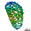
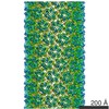
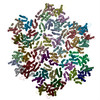
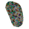
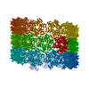
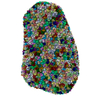
| タイトル | Atomic-level structure of the entire HIV-1 capsid (186 hexamers + 12 pentamers) | |||||||||
 要素 要素 | capsid protein | |||||||||
 キーワード キーワード | VIRUS / HIV-1 capsid / core / all-atom model / complete capsid | |||||||||
| 機能・相同性 |  機能・相同性情報 機能・相同性情報viral budding via host ESCRT complex / host multivesicular body / viral nucleocapsid / viral translational frameshifting / host cell nucleus / host cell plasma membrane / virion membrane / structural molecule activity / RNA binding / zinc ion binding ...viral budding via host ESCRT complex / host multivesicular body / viral nucleocapsid / viral translational frameshifting / host cell nucleus / host cell plasma membrane / virion membrane / structural molecule activity / RNA binding / zinc ion binding / identical protein binding / membrane 類似検索 - 分子機能 | |||||||||
| 生物種 |   Human immunodeficiency virus 1 (ヒト免疫不全ウイルス) Human immunodeficiency virus 1 (ヒト免疫不全ウイルス) | |||||||||
| 手法 | 電子顕微鏡法 / 電子線トモグラフィー法 / クライオ電子顕微鏡法 | |||||||||
 データ登録者 データ登録者 | Perilla, J.R. / Zhao, G. / Zhang, P. / Schulten, K.J. | |||||||||
 引用 引用 | ジャーナル: Nature / 年: 2013 タイトル: Mature HIV-1 capsid structure by cryo-electron microscopy and all-atom molecular dynamics. 著者: Gongpu Zhao / Juan R Perilla / Ernest L Yufenyuy / Xin Meng / Bo Chen / Jiying Ning / Jinwoo Ahn / Angela M Gronenborn / Klaus Schulten / Christopher Aiken / Peijun Zhang /  要旨: Retroviral capsid proteins are conserved structurally but assemble into different morphologies. The mature human immunodeficiency virus-1 (HIV-1) capsid is best described by a 'fullerene cone' model, ...Retroviral capsid proteins are conserved structurally but assemble into different morphologies. The mature human immunodeficiency virus-1 (HIV-1) capsid is best described by a 'fullerene cone' model, in which hexamers of the capsid protein are linked to form a hexagonal surface lattice that is closed by incorporating 12 capsid-protein pentamers. HIV-1 capsid protein contains an amino-terminal domain (NTD) comprising seven α-helices and a β-hairpin, a carboxy-terminal domain (CTD) comprising four α-helices, and a flexible linker with a 310-helix connecting the two structural domains. Structures of the capsid-protein assembly units have been determined by X-ray crystallography; however, structural information regarding the assembled capsid and the contacts between the assembly units is incomplete. Here we report the cryo-electron microscopy structure of a tubular HIV-1 capsid-protein assembly at 8 Å resolution and the three-dimensional structure of a native HIV-1 core by cryo-electron tomography. The structure of the tubular assembly shows, at the three-fold interface, a three-helix bundle with critical hydrophobic interactions. Mutagenesis studies confirm that hydrophobic residues in the centre of the three-helix bundle are crucial for capsid assembly and stability, and for viral infectivity. The cryo-electron-microscopy structures enable modelling by large-scale molecular dynamics simulation, resulting in all-atom models for the hexamer-of-hexamer and pentamer-of-hexamer elements as well as for the entire capsid. Incorporation of pentamers results in closer trimer contacts and induces acute surface curvature. The complete atomic HIV-1 capsid model provides a platform for further studies of capsid function and for targeted pharmacological intervention. | |||||||||
| 履歴 |
|
- 構造の表示
構造の表示
| ムービー |
ムービービューア |
|---|---|
| 構造ビューア | 分子: MolmilJmol/JSmol |
- ダウンロードとリンク
-ダウンロード
| PDBx/mmCIF形式 | 3j3y.cif.gz | 41.9 MB | 表示 | PDBx/mmCIF形式 |
|---|---|---|---|---|
| PDB形式 | pdb3j3y.ent.gz | 表示 | PDB形式 | |
| PDBx/mmJSON形式 | 3j3y.json.gz | ツリー表示 | PDBx/mmJSON形式 | |
| その他 |  その他のダウンロード その他のダウンロード |
-検証レポート
| 文書・要旨 | 3j3y_validation.pdf.gz | 3 MB | 表示 | wwPDB検証レポート |
|---|---|---|---|---|
| 文書・詳細版 | 3j3y_full_validation.pdf.gz | 4.5 MB | 表示 | |
| XML形式データ | 3j3y_validation.xml.gz | 2.9 MB | 表示 | |
| CIF形式データ | 3j3y_validation.cif.gz | 6.5 MB | 表示 | |
| アーカイブディレクトリ | https://data.pdbj.org/pub/pdb/validation_reports/j3/3j3yftp://data.pdbj.org/pub/pdb/validation_reports/j3/3j3y | HTTPS FTP |
-関連構造データ
-リンク
 PDBj
PDBj

- 集合体
集合体
| 登録構造単位 | 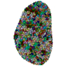
|
|---|---|
| 1 |
|
-要素
| #1: タンパク質 | 分子量: 25702.490 Da / 分子数: 1176 / 断片: UNP residues 133-363 / 変異: A92E / 由来タイプ: 組換発現 由来: (組換発現) Human immunodeficiency virus 1 (ヒト免疫不全ウイルス)遺伝子: gag / プラスミド: pET21 / 発現宿主:  Has protein modification | Y | |
|---|
-実験情報
-実験
| 実験 | 手法: 電子顕微鏡法 |
|---|---|
| EM実験 | 試料の集合状態: PARTICLE / 3次元再構成法: 電子線トモグラフィー法 |
- 試料調製
試料調製
| 構成要素 | 名称: HIV-1 core with A14C/E45C cross-linked capsid protein タイプ: VIRUS |
|---|---|
| ウイルスについての詳細 | 中空か: NO / エンベロープを持つか: YES / ホストのカテゴリ: VERTEBRATES / 単離: STRAIN / タイプ: VIRION |
| 天然宿主 | 生物種: Homo sapiens |
| 緩衝液 | 名称: 10 mM Tris-HCl, 100 mM NaCl, 1 mM EDTA / pH: 8 / 詳細: 10 mM Tris-HCl, 100 mM NaCl, 1 mM EDTA |
| 試料 | 濃度: 0.011 mg/ml / 包埋: NO / シャドウイング: NO / 染色: NO / 凍結: YES |
| 試料支持 | 詳細: Quantifoil R2/2 200 mesh holey carbon copper grids |
| 急速凍結 | 装置: HOMEMADE PLUNGER / 凍結剤: ETHANE |
- 電子顕微鏡撮影
| 実験機器 |  モデル: Tecnai Polara / 画像提供: FEI Company |
|---|---|
| 顕微鏡 | モデル: FEI POLARA 300 / 日付: 2010年9月11日 |
| 電子銃 | 電子線源:  FIELD EMISSION GUN / 加速電圧: 300 kV / 照射モード: FLOOD BEAM FIELD EMISSION GUN / 加速電圧: 300 kV / 照射モード: FLOOD BEAM |
| 電子レンズ | モード: BRIGHT FIELD / 倍率(公称値): 39000 X / 最大 デフォーカス(公称値): 8000 nm / 最小 デフォーカス(公称値): 8000 nm / Cs: 2 mm |
| 試料ホルダ | 試料ホルダーモデル: SIDE ENTRY, EUCENTRIC / 温度: 82 K / 最高温度: 85 K / 最低温度: 80 K / 傾斜角・最大: 66 ° / 傾斜角・最小: -70 ° |
| 撮影 | 電子線照射量: 120 e/Å2 フィルム・検出器のモデル: GATAN ULTRASCAN 4000 (4k x 4k) |
| 画像スキャン | デジタル画像の数: 53 |
- 解析
解析
| EMソフトウェア |
| |||||||||
|---|---|---|---|---|---|---|---|---|---|---|
| 対称性 | 点対称性: C1 (非対称) | |||||||||
| 3次元再構成 | 手法: SIRT / ピクセルサイズ(公称値): 6.25 Å / ピクセルサイズ(実測値): 6.25 Å / 対称性のタイプ: POINT | |||||||||
| 原子モデル構築 | プロトコル: FLEXIBLE FIT / 空間: REAL 詳細: REFINEMENT PROTOCOL--FLEXIBLE DETAILS--The model was built using hexamer of hexamers (PDB entry 3J34) and pentamer of hexamers (computer-based MD model available upon request). | |||||||||
| 原子モデル構築 | PDB-ID: 3J34 Accession code: 3J34 / Source name: PDB / タイプ: experimental model |
