 Movie
Movie Controller


+ Open data

- Basic information
| Entry | Database: PDB / ID: 3ip4 | ||||||
|---|---|---|---|---|---|---|---|
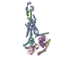
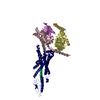
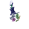
| Title | The high resolution structure of GatCAB | ||||||
 Components Components |
| ||||||
 Keywords Keywords | LIGASE / MULTI PROTEIN COMPLEX / ATP-binding / Nucleotide-binding / Protein biosynthesis | ||||||
| Function / homology |  Function and homology information Function and homology informationasparaginyl-tRNA synthase (glutamine-hydrolyzing) activity / glutaminyl-tRNA synthase (glutamine-hydrolysing) / glutamyl-tRNA(Gln) amidotransferase complex / Ligases; Forming carbon-nitrogen bonds; Carbon-nitrogen ligases with glutamine as amido-N-donor / glutaminyl-tRNAGln biosynthesis via transamidation / glutaminyl-tRNA synthase (glutamine-hydrolyzing) activity / regulation of translational fidelity / translation / ATP binding Similarity search - Function | ||||||
| Biological species |   Staphylococcus aureus subsp. aureus (bacteria) Staphylococcus aureus subsp. aureus (bacteria) | ||||||
| Method |  X-RAY DIFFRACTION / SYNCHROTRON / MOLECULAR REPLACEMENT / Resolution: 1.9 Å X-RAY DIFFRACTION / SYNCHROTRON / MOLECULAR REPLACEMENT / Resolution: 1.9 Å | ||||||
 Authors Authors | Nakamura, A. / Yao, M. / Tanaka, I. | ||||||
 Citation Citation | Journal: Nucleic Acids Res. / Year: 2010 Title: Two distinct regions in Staphylococcus aureus GatCAB guarantee accurate tRNA recognition Authors: Nakamura, A. / Sheppard, K. / Yamane, J. / Yao, M. / Soll, D. / Tanaka, I. | ||||||
| History |
|
- Structure visualization
| Structure viewer | Molecule: MolmilJmol/JSmol |
|---|
- Downloads & links
-Download
| PDBx/mmCIF format | 3ip4.cif.gz | 235.9 KB | Display | PDBx/mmCIF format |
|---|---|---|---|---|
| PDB format | pdb3ip4.ent.gz | 184.2 KB | Display | PDB format |
| PDBx/mmJSON format | 3ip4.json.gz | Tree view | PDBx/mmJSON format | |
| Others |  Other downloads Other downloads |
-Validation report
| Arichive directory | https://data.pdbj.org/pub/pdb/validation_reports/ip/3ip4ftp://data.pdbj.org/pub/pdb/validation_reports/ip/3ip4 | HTTPS FTP |
|---|
-Related structure data
| Related structure data | 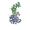 2g5hS S: Starting model for refinement |
|---|---|
| Similar structure data |


-Links
 PDBj
PDBj
- Assembly
| Deposited unit | 
| ||||||||
|---|---|---|---|---|---|---|---|---|---|
| 1 |
| ||||||||
| Unit cell |
|
-Components
| #1: Protein | Mass: 52858.660 Da / Num. of mol.: 1 Source method: isolated from a genetically manipulated source Source: (gene. exp.) Staphylococcus aureus subsp. aureus (bacteria)Strain: Mu50 / Gene: gatA / Plasmid: pET28b / Production host: References: UniProt: P63488, Ligases; Forming carbon-nitrogen bonds; Carbon-nitrogen ligases with glutamine as amido-N-donor |
|---|---|
| #2: Protein | Mass: 54794.633 Da / Num. of mol.: 1 Source method: isolated from a genetically manipulated source Source: (gene. exp.) Staphylococcus aureus subsp. aureus (bacteria)Strain: Mu50 / Gene: gatB / Plasmid: pET28b / Production host: References: UniProt: P64201, Ligases; Forming carbon-nitrogen bonds; Carbon-nitrogen ligases with glutamine as amido-N-donor |
| #3: Protein | Mass: 11279.481 Da / Num. of mol.: 1 Source method: isolated from a genetically manipulated source Source: (gene. exp.) Staphylococcus aureus subsp. aureus (bacteria)Strain: Mu50 / Gene: gatC / Plasmid: pET28b / Production host: References: UniProt: P68807, Ligases; Forming carbon-nitrogen bonds; Carbon-nitrogen ligases with glutamine as amido-N-donor |
| #4: Chemical | ChemComp-MG /   Mass: 24.305 Da / Num. of mol.: 1 / Source method: obtained synthetically / Formula: Mg Mass: 24.305 Da / Num. of mol.: 1 / Source method: obtained synthetically / Formula: Mg |
| #5: Water | ChemComp-HOH /  Mass: 18.015 Da / Num. of mol.: 801 / Source method: isolated from a natural source / Formula: H2O Mass: 18.015 Da / Num. of mol.: 801 / Source method: isolated from a natural source / Formula: H2O |
-Experimental details
-Experiment
| Experiment | Method: X-RAY DIFFRACTION / Number of used crystals: 1 |
|---|
- Sample preparation
| Crystal | Density Matthews: 2.5 Å3/Da / Density % sol: 50.8 % |
|---|---|
| Crystal grow | Temperature: 293 K / Method: vapor diffusion, hanging drop / pH: 7.2 Details: 25% PEG 600, 5mM magnesium chloride, 50mM HEPES-NaOH, 3% MPD, pH 7.2, VAPOR DIFFUSION, HANGING DROP, temperature 293K |
-Data collection
| Diffraction | Mean temperature: 100 K |
|---|---|
| Diffraction source | Source: SYNCHROTRON / Site: SPring-8  / Beamline: BL41XU / Wavelength: 1 Å / Beamline: BL41XU / Wavelength: 1 Å |
| Detector | Type: ADSC QUANTUM 315 / Detector: CCD / Date: Jun 12, 2007 / Details: mirrors |
| Radiation | Monochromator: MIRRORS / Protocol: SINGLE WAVELENGTH / Monochromatic (M) / Laue (L): M / Scattering type: x-ray |
| Radiation wavelength | Wavelength: 1 Å / Relative weight: 1 |
| Reflection | Resolution: 1.9→50 Å / Num. obs: 94418 / % possible obs: 99.6 % / Observed criterion σ(F): 0 / Observed criterion σ(I): -3 / Redundancy: 7 % / Biso Wilson estimate: 25.7 Å2 / Rmerge(I) obs: 0.07 / Rsym value: 0.07 / Net I/σ(I): 23.2 |
| Reflection shell | Resolution: 1.9→1.97 Å / Redundancy: 5.5 % / Rmerge(I) obs: 0.457 / Mean I/σ(I) obs: 3.5 / Num. unique all: 9320 / Rsym value: 0.457 / % possible all: 100 |
- Processing
| Software |
| ||||||||||||||||||||
|---|---|---|---|---|---|---|---|---|---|---|---|---|---|---|---|---|---|---|---|---|---|
| Refinement | Method to determine structure: MOLECULAR REPLACEMENT Starting model: PDB ENTRY 2G5H Resolution: 1.9→20 Å / Isotropic thermal model: Isotropic / Cross valid method: THROUGHOUT / σ(F): 0 / σ(I): 0 / Stereochemistry target values: Engh & Huber
| ||||||||||||||||||||
| Displacement parameters | Biso mean: 49.9 Å2
| ||||||||||||||||||||
| Refine analyze |
| ||||||||||||||||||||
| Refinement step | Cycle: LAST / Resolution: 1.9→20 Å
| ||||||||||||||||||||
| Refine LS restraints |
| ||||||||||||||||||||
| LS refinement shell | Resolution: 1.9→1.97 Å
|
