 Movie
Movie Controller

[English] 日本語
 Yorodumi
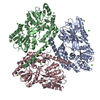
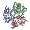
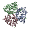
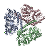
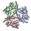
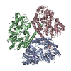
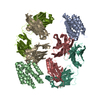
- PDB-3ior: Huntingtin amino-terminal region with 17 Gln residues - crystal C95 -
+ Open data

- Basic information
| Entry | Database: PDB / ID: 3ior | ||||||
|---|---|---|---|---|---|---|---|
| Title | Huntingtin amino-terminal region with 17 Gln residues - crystal C95 | ||||||
 Components Components | Maltose-binding protein, huntingtin fusion protein | ||||||
 Keywords Keywords | SIGNALING PROTEIN / Huntingtin / Htt-Ex1 / HD / Sugar transport / Transport / Apoptosis / Disease mutation / Nucleus / Phosphoprotein | ||||||
| Function / homology |  Function and homology information Function and homology informationpositive regulation of CAMKK-AMPK signaling cascade / vocal learning / regulation of CAMKK-AMPK signaling cascade / positive regulation of mitophagy / profilin binding / positive regulation of cilium assembly / retrograde vesicle-mediated transport, Golgi to endoplasmic reticulum / vesicle transport along microtubule / positive regulation of aggrephagy / positive regulation of lipophagy ...positive regulation of CAMKK-AMPK signaling cascade / vocal learning / regulation of CAMKK-AMPK signaling cascade / positive regulation of mitophagy / profilin binding / positive regulation of cilium assembly / retrograde vesicle-mediated transport, Golgi to endoplasmic reticulum / vesicle transport along microtubule / positive regulation of aggrephagy / positive regulation of lipophagy / Golgi organization / establishment of mitotic spindle orientation / detection of maltose stimulus / dynein intermediate chain binding / maltose transport complex / synaptic vesicle transport / carbohydrate transport / dynactin binding / Regulation of MECP2 expression and activity / carbohydrate transmembrane transporter activity / maltose binding / maltose transport / maltodextrin transmembrane transport / postsynaptic cytosol / beta-tubulin binding / presynaptic cytosol / ATP-binding cassette (ABC) transporter complex, substrate-binding subunit-containing / positive regulation of calcium-mediated signaling / phosphoprotein phosphatase activity / heat shock protein binding / neurogenesis / centriole / inclusion body / autophagosome / ATP-binding cassette (ABC) transporter complex / cytoplasmic vesicle membrane / negative regulation of extrinsic apoptotic signaling pathway / central nervous system development / cell chemotaxis / protein destabilization / kinase binding / p53 binding / late endosome / outer membrane-bounded periplasmic space / cytoplasmic vesicle / transmembrane transporter binding / early endosome / periplasmic space / positive regulation of apoptotic process / axon / apoptotic process / DNA damage response / dendrite / perinuclear region of cytoplasm / Golgi apparatus / endoplasmic reticulum / protein-containing complex / nucleoplasm / membrane / identical protein binding / nucleus / cytosol / cytoplasm Similarity search - Function | ||||||
| Biological species |   Homo sapiens (human) Homo sapiens (human) | ||||||
| Method |  X-RAY DIFFRACTION / SYNCHROTRON / MOLECULAR REPLACEMENT / Resolution: 3.6 Å X-RAY DIFFRACTION / SYNCHROTRON / MOLECULAR REPLACEMENT / Resolution: 3.6 Å | ||||||
 Authors Authors | Kim, M.W. | ||||||
 Citation Citation | Journal: Structure / Year: 2009 Title: Secondary structure of Huntingtin amino-terminal region. Authors: Kim, M.W. / Chelliah, Y. / Kim, S.W. / Otwinowski, Z. / Bezprozvanny, I. | ||||||
| History |
|
- Structure visualization
| Structure viewer | Molecule: MolmilJmol/JSmol |
|---|
- Downloads & links
-Download
| PDBx/mmCIF format | 3ior.cif.gz | 423.3 KB | Display | PDBx/mmCIF format |
|---|---|---|---|---|
| PDB format | pdb3ior.ent.gz | 346.2 KB | Display | PDB format |
| PDBx/mmJSON format | 3ior.json.gz | Tree view | PDBx/mmJSON format | |
| Others |  Other downloads Other downloads |
-Validation report
| Arichive directory | https://data.pdbj.org/pub/pdb/validation_reports/io/3iorftp://data.pdbj.org/pub/pdb/validation_reports/io/3ior | HTTPS FTP |
|---|
-Related structure data
| Related structure data | 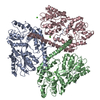 3io4C  3io6C  3iotC  3iouC  3iovC  3iowC C: citing same article ( |
|---|---|
| Similar structure data |


-Links
 PDBj
PDBj


- Assembly
| Deposited unit | 
| ||||||||
|---|---|---|---|---|---|---|---|---|---|
| 1 |
| ||||||||
| Unit cell |
| ||||||||
| Details | AUTHORS STATE THAT THE BIOLOGICAL UNIT OF THIS POLYPEPTIDE IS UNKNOWN. |
-Components
| #1: Protein | Mass: 49316.715 Da / Num. of mol.: 3 / Fragment: Fusion protein, see remark 999 Source method: isolated from a genetically manipulated source Details: Huntingtin-Ex1 / Source: (gene. exp.) Escherichia coli K-12, Homo sapiens / Gene: malE, b4034, JW3994, HTT, HD, IT15 / Plasmid: pMAL / Production host: #2: Chemical | ChemComp-ZN /   Mass: 65.409 Da / Num. of mol.: 6 / Source method: obtained synthetically / Formula: Zn Mass: 65.409 Da / Num. of mol.: 6 / Source method: obtained synthetically / Formula: Zn#3: Chemical | ChemComp-CA /   Mass: 40.078 Da / Num. of mol.: 6 / Source method: obtained synthetically / Formula: Ca Mass: 40.078 Da / Num. of mol.: 6 / Source method: obtained synthetically / Formula: CaSequence details | ENTITY 1 IS A FUSION PROTEIN OF E.COLI MALTOSE BINDING PROTEIN (UNIPROT P0AEX9 (MALE_ECOLI) ...ENTITY 1 IS A FUSION PROTEIN OF E.COLI MALTOSE BINDING PROTEIN (UNIPROT P0AEX9 (MALE_ECOLI) RESIDUES 27-384) TO N-TERMINAL RESIDUES 1-64 OF HUMAN HUNTINGTIN | |
|---|
-Experimental details
-Experiment
| Experiment | Method: X-RAY DIFFRACTION / Number of used crystals: 1 |
|---|
- Sample preparation
| Crystal | Density Matthews: 3.96 Å3/Da / Density % sol: 68.96 % |
|---|---|
| Crystal grow | Temperature: 278 K / Method: vapor diffusion, hanging drop Details: 12% PEG 4000, 200 mM Zn acetate, 200 mM Sodium acetate, 100 mM Sodium Cacodylate pH 6.5-7.4 , VAPOR DIFFUSION, HANGING DROP, temperature 278K PH range: 6.5-7.4 |
-Data collection
| Diffraction | Mean temperature: 100 K |
|---|---|
| Diffraction source | Source: SYNCHROTRON / Site: APS  / Beamline: 19-ID / Wavelength: 0.98 Å / Beamline: 19-ID / Wavelength: 0.98 Å |
| Detector | Type: ADSC QUANTUM 315 / Detector: CCD Details: LN2 cooled first crystal, sagitally focusing 2nd crystal, Rosenbaum-Rock vertical focusing mirror, beam defining slits |
| Radiation | Monochromator: Rosenbaum-Rock high-resolution Si(111) double-crystal Protocol: SINGLE WAVELENGTH / Monochromatic (M) / Laue (L): M / Scattering type: x-ray |
| Radiation wavelength | Wavelength: 0.98 Å / Relative weight: 1 |
| Reflection | Resolution: 2.68→50 Å / Num. obs: 20472 / Redundancy: 2.5 % / Rsym value: 0.09 |
- Processing
| Software |
| ||||||||||||||||||||||||||||||||||||||||||||||||||||||||||||||||||||||||||||||||||||||||||||||||||||||||||||||||||||||||||||||||||||||||||||||||||||||||||||||||||||||||||
|---|---|---|---|---|---|---|---|---|---|---|---|---|---|---|---|---|---|---|---|---|---|---|---|---|---|---|---|---|---|---|---|---|---|---|---|---|---|---|---|---|---|---|---|---|---|---|---|---|---|---|---|---|---|---|---|---|---|---|---|---|---|---|---|---|---|---|---|---|---|---|---|---|---|---|---|---|---|---|---|---|---|---|---|---|---|---|---|---|---|---|---|---|---|---|---|---|---|---|---|---|---|---|---|---|---|---|---|---|---|---|---|---|---|---|---|---|---|---|---|---|---|---|---|---|---|---|---|---|---|---|---|---|---|---|---|---|---|---|---|---|---|---|---|---|---|---|---|---|---|---|---|---|---|---|---|---|---|---|---|---|---|---|---|---|---|---|---|---|---|---|---|
| Refinement | Method to determine structure: MOLECULAR REPLACEMENT / Resolution: 3.6→37.36 Å / Cor.coef. Fo:Fc: 0.853 / Cor.coef. Fo:Fc free: 0.857 / SU B: 68.196 / SU ML: 0.461 / Cross valid method: THROUGHOUT / ESU R Free: 0.697 / Stereochemistry target values: MAXIMUM LIKELIHOOD Details: HYDROGENS HAVE BEEN ADDED IN THE RIDING POSITIONS. U VALUES: REFINED INDIVIDUALLY. AUTHORS USED NCS RESTRAINTS IN REFINEMENT.
| ||||||||||||||||||||||||||||||||||||||||||||||||||||||||||||||||||||||||||||||||||||||||||||||||||||||||||||||||||||||||||||||||||||||||||||||||||||||||||||||||||||||||||
| Solvent computation | Ion probe radii: 0.8 Å / Shrinkage radii: 0.8 Å / VDW probe radii: 1.4 Å / Solvent model: MASK | ||||||||||||||||||||||||||||||||||||||||||||||||||||||||||||||||||||||||||||||||||||||||||||||||||||||||||||||||||||||||||||||||||||||||||||||||||||||||||||||||||||||||||
| Displacement parameters | Biso mean: 56.475 Å2
| ||||||||||||||||||||||||||||||||||||||||||||||||||||||||||||||||||||||||||||||||||||||||||||||||||||||||||||||||||||||||||||||||||||||||||||||||||||||||||||||||||||||||||
| Refinement step | Cycle: LAST / Resolution: 3.6→37.36 Å
| ||||||||||||||||||||||||||||||||||||||||||||||||||||||||||||||||||||||||||||||||||||||||||||||||||||||||||||||||||||||||||||||||||||||||||||||||||||||||||||||||||||||||||
| Refine LS restraints |
| ||||||||||||||||||||||||||||||||||||||||||||||||||||||||||||||||||||||||||||||||||||||||||||||||||||||||||||||||||||||||||||||||||||||||||||||||||||||||||||||||||||||||||
| LS refinement shell | Resolution: 3.6→3.693 Å / Total num. of bins used: 20
|
