 Movie
Movie Controller


+ Open data

- Basic information
| Entry | Database: PDB / ID: 3iig | ||||||
|---|---|---|---|---|---|---|---|
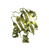
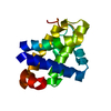
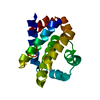
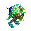
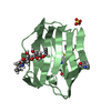
| Title | Crystal structure of mouse Bcl-xl mutant (F105A) at pH 5.0 | ||||||
 Components Components | Bcl-2-like protein 1 | ||||||
 Keywords Keywords | APOPTOSIS / BH3 domain / Bcl-2 / Membrane / Mitochondrion / Transmembrane | ||||||
| Function / homology |  Function and homology information Function and homology informationThe NLRP1 inflammasome / BH3-only proteins associate with and inactivate anti-apoptotic BCL-2 members / synaptic vesicle recycling via endosome / positive regulation of synaptic vesicle exocytosis / positive regulation of synaptic vesicle clustering / RAS processing / BH domain binding / apoptotic process in bone marrow cell / positive regulation of synaptic vesicle endocytosis / negative regulation of intrinsic apoptotic signaling pathway in response to DNA damage ...The NLRP1 inflammasome / BH3-only proteins associate with and inactivate anti-apoptotic BCL-2 members / synaptic vesicle recycling via endosome / positive regulation of synaptic vesicle exocytosis / positive regulation of synaptic vesicle clustering / RAS processing / BH domain binding / apoptotic process in bone marrow cell / positive regulation of synaptic vesicle endocytosis / negative regulation of intrinsic apoptotic signaling pathway in response to DNA damage / negative regulation of mitochondrial outer membrane permeabilization involved in apoptotic signaling pathway / cysteine-type endopeptidase inhibitor activity involved in apoptotic process / negative regulation of execution phase of apoptosis / fertilization / regulation of mitochondrial membrane permeability / regulation of growth / Bcl-2 family protein complex / response to cycloheximide / clathrin binding / negative regulation of release of cytochrome c from mitochondria / cellular response to alkaloid / hepatocyte apoptotic process / negative regulation of intrinsic apoptotic signaling pathway / germ cell development / positive regulation of ATP biosynthetic process / negative regulation of anoikis / BH3 domain binding / regulation of synaptic vesicle endocytosis / negative regulation of protein localization to plasma membrane / negative regulation of endoplasmic reticulum stress-induced intrinsic apoptotic signaling pathway / negative regulation of extrinsic apoptotic signaling pathway via death domain receptors / ovarian follicle development / extrinsic apoptotic signaling pathway in absence of ligand / regulation of long-term synaptic depression / clathrin-coated pit / response to cytokine / MDM2/MDM4 family protein binding / release of cytochrome c from mitochondria / response to ischemia / regulation of cytokinesis / regulation of mitochondrial membrane potential / mitochondrion organization / cellular response to amino acid stimulus / cellular response to gamma radiation / response to radiation / male gonad development / mitochondrial membrane / intrinsic apoptotic signaling pathway in response to DNA damage / response to virus / synaptic vesicle / synaptic vesicle membrane / presynapse / channel activity / GTPase binding / neuron apoptotic process / spermatogenesis / regulation of apoptotic process / nuclear membrane / defense response to virus / in utero embryonic development / negative regulation of neuron apoptotic process / mitochondrial outer membrane / cell population proliferation / mitochondrial inner membrane / positive regulation of apoptotic process / mitochondrial matrix / positive regulation of cell population proliferation / centrosome / protein kinase binding / negative regulation of apoptotic process / protein-containing complex binding / endoplasmic reticulum / mitochondrion / membrane / identical protein binding / cytoplasm / cytosol Similarity search - Function | ||||||
| Biological species |  | ||||||
| Method |  X-RAY DIFFRACTION / SYNCHROTRON / MOLECULAR REPLACEMENT / Resolution: 2.3 Å X-RAY DIFFRACTION / SYNCHROTRON / MOLECULAR REPLACEMENT / Resolution: 2.3 Å | ||||||
 Authors Authors | Priyadarshi, A. / Hwang, K.Y. | ||||||
 Citation Citation | Journal: Biochem.Biophys.Res.Commun. / Year: 2010 Title: Structural insights into mouse anti-apoptotic Bcl-xl reveal affinity for Beclin 1 and gossypol. Authors: Priyadarshi, A. / Roy, A. / Kim, K.S. / Kim, E.E. / Hwang, K.Y. | ||||||
| History |
|
- Structure visualization
| Structure viewer | Molecule: MolmilJmol/JSmol |
|---|
- Downloads & links
-Download
| PDBx/mmCIF format | 3iig.cif.gz | 42.1 KB | Display | PDBx/mmCIF format |
|---|---|---|---|---|
| PDB format | pdb3iig.ent.gz | 29 KB | Display | PDB format |
| PDBx/mmJSON format | 3iig.json.gz | Tree view | PDBx/mmJSON format | |
| Others |  Other downloads Other downloads |
-Validation report
| Arichive directory | https://data.pdbj.org/pub/pdb/validation_reports/ii/3iigftp://data.pdbj.org/pub/pdb/validation_reports/ii/3iig | HTTPS FTP |
|---|
-Related structure data
| Related structure data |  3ihcC  3ihdC  3iheC  3ihfC  3iihC  3ilbC  3ilcC  1pq0S C: citing same article ( S: Starting model for refinement |
|---|---|
| Similar structure data |

-Links
 PDBj
PDBj
- Assembly
| Deposited unit | 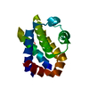
| ||||||||
|---|---|---|---|---|---|---|---|---|---|
| 1 |
| ||||||||
| Unit cell |
|
-Components
| #1: Protein | Mass: 21980.113 Da / Num. of mol.: 1 / Fragment: UNP residues 1-196 / Mutation: F105A Source method: isolated from a genetically manipulated source Source: (gene. exp.)  |
|---|---|
| #2: Water | ChemComp-HOH /  Mass: 18.015 Da / Num. of mol.: 11 / Source method: isolated from a natural source / Formula: H2O Mass: 18.015 Da / Num. of mol.: 11 / Source method: isolated from a natural source / Formula: H2O |
-Experimental details
-Experiment
| Experiment | Method: X-RAY DIFFRACTION / Number of used crystals: 1 |
|---|
- Sample preparation
| Crystal | Density Matthews: 2.5 Å3/Da / Density % sol: 50.86 % |
|---|---|
| Crystal grow | Temperature: 295 K / Method: vapor diffusion, hanging drop / pH: 5 Details: 1.6M Ammonium sulphate, 0.1M tri-Na citrate, pH 5.0, VAPOR DIFFUSION, HANGING DROP, temperature 295K |
-Data collection
| Diffraction | Mean temperature: 100 K |
|---|---|
| Diffraction source | Source: SYNCHROTRON / Site: PAL/PLS  / Beamline: 6C1 / Wavelength: 1 Å / Beamline: 6C1 / Wavelength: 1 Å |
| Detector | Type: ADSC QUANTUM 270 / Detector: CCD / Date: Oct 8, 2008 / Details: mirrors |
| Radiation | Monochromator: Graphite / Protocol: SINGLE WAVELENGTH / Monochromatic (M) / Laue (L): M / Scattering type: x-ray |
| Radiation wavelength | Wavelength: 1 Å / Relative weight: 1 |
| Reflection | Resolution: 2.3→40 Å / Num. all: 9693 / Num. obs: 9215 / % possible obs: 92.4 % / Observed criterion σ(F): 0 / Observed criterion σ(I): 0 / Redundancy: 8.6 % / Biso Wilson estimate: 48.8 Å2 / Rmerge(I) obs: 0.15 / Rsym value: 0.24 / Net I/σ(I): 20.5 |
| Reflection shell | Resolution: 2.3→2.38 Å / Redundancy: 4.2 % / Rmerge(I) obs: 0.15 / Mean I/σ(I) obs: 3.1 / Num. unique all: 772 / Rsym value: 0.24 / % possible all: 76.4 |
- Processing
| Software |
| ||||||||||||||||||||||||||||||||||||||||||||||||||||||||||||||||||||||||||||||||||||||||||
|---|---|---|---|---|---|---|---|---|---|---|---|---|---|---|---|---|---|---|---|---|---|---|---|---|---|---|---|---|---|---|---|---|---|---|---|---|---|---|---|---|---|---|---|---|---|---|---|---|---|---|---|---|---|---|---|---|---|---|---|---|---|---|---|---|---|---|---|---|---|---|---|---|---|---|---|---|---|---|---|---|---|---|---|---|---|---|---|---|---|---|---|
| Refinement | Method to determine structure: MOLECULAR REPLACEMENT Starting model: 1PQ0 Resolution: 2.3→40 Å / Cor.coef. Fo:Fc: 0.918 / Cor.coef. Fo:Fc free: 0.903 / SU B: 6.739 / SU ML: 0.166 / Cross valid method: THROUGHOUT / σ(I): 0 / ESU R: 0.283 / ESU R Free: 0.228 / Stereochemistry target values: MAXIMUM LIKELIHOOD / Details: HYDROGENS HAVE BEEN ADDED IN THE RIDING POSITIONS
| ||||||||||||||||||||||||||||||||||||||||||||||||||||||||||||||||||||||||||||||||||||||||||
| Solvent computation | Ion probe radii: 0.8 Å / Shrinkage radii: 0.8 Å / VDW probe radii: 1.4 Å / Solvent model: MASK | ||||||||||||||||||||||||||||||||||||||||||||||||||||||||||||||||||||||||||||||||||||||||||
| Displacement parameters | Biso mean: 48.869 Å2
| ||||||||||||||||||||||||||||||||||||||||||||||||||||||||||||||||||||||||||||||||||||||||||
| Refinement step | Cycle: LAST / Resolution: 2.3→40 Å
| ||||||||||||||||||||||||||||||||||||||||||||||||||||||||||||||||||||||||||||||||||||||||||
| Refine LS restraints |
| ||||||||||||||||||||||||||||||||||||||||||||||||||||||||||||||||||||||||||||||||||||||||||
| LS refinement shell | Resolution: 2.3→2.36 Å / Total num. of bins used: 20
|
