 Movie
Movie Controller

[English] 日本語
 Yorodumi
- PDB-3igm: A 2.2A crystal structure of the AP2 domain of PF14_0633 from P. f... -
+ Open data

- Basic information
| Entry | Database: PDB / ID: 3igm | ||||||
|---|---|---|---|---|---|---|---|
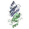
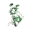
| Title | A 2.2A crystal structure of the AP2 domain of PF14_0633 from P. falciparum, bound as a domain-swapped dimer to its cognate DNA | ||||||
 Components Components |
| ||||||
 Keywords Keywords | Transcription/DNA / AP2 domain / Plasmodium falciparum / specific transcription factor / PROTEIN-DNA COMPLEX / Transcription-DNA COMPLEX | ||||||
| Function / homology |  Function and homology information Function and homology informationsequence-specific DNA binding / DNA-binding transcription factor activity / nucleus Similarity search - Function | ||||||
| Biological species |  | ||||||
| Method |  X-RAY DIFFRACTION / SYNCHROTRON / SAD / Resolution: 2.2 Å X-RAY DIFFRACTION / SYNCHROTRON / SAD / Resolution: 2.2 Å | ||||||
 Authors Authors | Lindner, S.E. / De Silva, E. / Keck, J.L. / Llinas, M. | ||||||
 Citation Citation | Journal: J.Mol.Biol. / Year: 2010 Title: Structural Determinants of DNA Binding by a P. falciparum ApiAP2 Transcriptional Regulator. Authors: Lindner, S.E. / De Silva, E.K. / Keck, J.L. / Llinas, M. | ||||||
| History |
|
- Structure visualization
| Structure viewer | Molecule: MolmilJmol/JSmol |
|---|
- Downloads & links
-Download
| PDBx/mmCIF format | 3igm.cif.gz | 59 KB | Display | PDBx/mmCIF format |
|---|---|---|---|---|
| PDB format | pdb3igm.ent.gz | 40.2 KB | Display | PDB format |
| PDBx/mmJSON format | 3igm.json.gz | Tree view | PDBx/mmJSON format | |
| Others |  Other downloads Other downloads |
-Validation report
| Arichive directory | https://data.pdbj.org/pub/pdb/validation_reports/ig/3igmftp://data.pdbj.org/pub/pdb/validation_reports/ig/3igm | HTTPS FTP |
|---|
-Related structure data
| Similar structure data |
|---|


-Links
 PDBj
PDBj- Assembly
| Deposited unit | 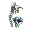
| ||||||||
|---|---|---|---|---|---|---|---|---|---|
| 1 |
| ||||||||
| Unit cell |
| ||||||||
| Details | BIOLOGICAL ASSEMBLY OF PROTEIN IS DIMER |
-Components
| #1: Protein | Mass: 8815.056 Da / Num. of mol.: 2 / Fragment: UNP residues 63-123, AP2 domain Source method: isolated from a genetically manipulated source Details: GST-tagged derivative of the AP2 domain of PF14_0633. GST removed by thrombin cleavage. Source: (gene. exp.) Strain: 3D7 / Gene: FP14_0633, PF14_0633 / Plasmid: pSURE, a pGEX4T-1 derivative / Production host:  #2: DNA chain | Mass: 2426.617 Da / Num. of mol.: 4 / Source method: obtained synthetically Details: This sequence occurs naturally in P. falciparum. DNA produced for this study was chemically synthesized. #3: Water | ChemComp-HOH / |  Mass: 18.015 Da / Num. of mol.: 97 / Source method: isolated from a natural source / Formula: H2O Mass: 18.015 Da / Num. of mol.: 97 / Source method: isolated from a natural source / Formula: H2OHas protein modification | Y | |
|---|
-Experimental details
-Experiment
| Experiment | Method: X-RAY DIFFRACTION / Number of used crystals: 1 |
|---|
- Sample preparation
| Crystal | Density Matthews: 2.09 Å3/Da / Density % sol: 41.12 % |
|---|---|
| Crystal grow | Temperature: 295 K / Method: vapor diffusion, hanging drop / pH: 5.1 Details: 0.5M-1.25M 1,6-hexanediol 100mM NaOAc pH 5.1 @295K 10mM CoCl2 10-20% ethylene glycol, VAPOR DIFFUSION, HANGING DROP |
-Data collection
| Diffraction | Mean temperature: 100 K |
|---|---|
| Diffraction source | Source: SYNCHROTRON / Site: APS  / Beamline: 21-ID-D / Wavelength: 0.97886 Å / Beamline: 21-ID-D / Wavelength: 0.97886 Å |
| Detector | Type: MARMOSAIC 300 mm CCD / Detector: CCD / Date: Dec 10, 2008 |
| Radiation | Monochromator: Si(111) / Protocol: SINGLE WAVELENGTH / Monochromatic (M) / Laue (L): M / Scattering type: x-ray |
| Radiation wavelength | Wavelength: 0.97886 Å / Relative weight: 1 |
| Reflection | Resolution: 2.2→50 Å / Num. all: 11402 / Num. obs: 10478 / % possible obs: 91.9 % / Observed criterion σ(F): 0 / Observed criterion σ(I): 0 / Redundancy: 11 % / Rsym value: 0.075 / Net I/σ(I): 32.1 |
| Reflection shell | Resolution: 2.2→2.26 Å / Redundancy: 9 % / Mean I/σ(I) obs: 4.5 / Rsym value: 0.381 / % possible all: 72.4 |
- Processing
| Software |
| ||||||||||||||||||||||||||||||||||||||||||||||||||||||||||||||||||||||||||||||||||||||||||||||||||||||||||||||||||||||||||||||||||||||||||||||||||||||||||||||||||||||||||
|---|---|---|---|---|---|---|---|---|---|---|---|---|---|---|---|---|---|---|---|---|---|---|---|---|---|---|---|---|---|---|---|---|---|---|---|---|---|---|---|---|---|---|---|---|---|---|---|---|---|---|---|---|---|---|---|---|---|---|---|---|---|---|---|---|---|---|---|---|---|---|---|---|---|---|---|---|---|---|---|---|---|---|---|---|---|---|---|---|---|---|---|---|---|---|---|---|---|---|---|---|---|---|---|---|---|---|---|---|---|---|---|---|---|---|---|---|---|---|---|---|---|---|---|---|---|---|---|---|---|---|---|---|---|---|---|---|---|---|---|---|---|---|---|---|---|---|---|---|---|---|---|---|---|---|---|---|---|---|---|---|---|---|---|---|---|---|---|---|---|---|---|
| Refinement | Method to determine structure: SAD / Resolution: 2.2→29.55 Å / Cor.coef. Fo:Fc: 0.949 / Cor.coef. Fo:Fc free: 0.925 / SU B: 15.931 / SU ML: 0.18 / TLS residual ADP flag: LIKELY RESIDUAL / Cross valid method: THROUGHOUT / ESU R: 0.334 / ESU R Free: 0.243 / Stereochemistry target values: MAXIMUM LIKELIHOOD / Details: HYDROGENS HAVE BEEN ADDED IN THE RIDING POSITIONS
| ||||||||||||||||||||||||||||||||||||||||||||||||||||||||||||||||||||||||||||||||||||||||||||||||||||||||||||||||||||||||||||||||||||||||||||||||||||||||||||||||||||||||||
| Solvent computation | Ion probe radii: 0.8 Å / Shrinkage radii: 0.8 Å / VDW probe radii: 1.4 Å / Solvent model: BABINET MODEL WITH MASK | ||||||||||||||||||||||||||||||||||||||||||||||||||||||||||||||||||||||||||||||||||||||||||||||||||||||||||||||||||||||||||||||||||||||||||||||||||||||||||||||||||||||||||
| Displacement parameters | Biso mean: 48.953 Å2
| ||||||||||||||||||||||||||||||||||||||||||||||||||||||||||||||||||||||||||||||||||||||||||||||||||||||||||||||||||||||||||||||||||||||||||||||||||||||||||||||||||||||||||
| Refinement step | Cycle: LAST / Resolution: 2.2→29.55 Å
| ||||||||||||||||||||||||||||||||||||||||||||||||||||||||||||||||||||||||||||||||||||||||||||||||||||||||||||||||||||||||||||||||||||||||||||||||||||||||||||||||||||||||||
| Refine LS restraints |
| ||||||||||||||||||||||||||||||||||||||||||||||||||||||||||||||||||||||||||||||||||||||||||||||||||||||||||||||||||||||||||||||||||||||||||||||||||||||||||||||||||||||||||
| LS refinement shell | Resolution: 2.2→2.262 Å / Total num. of bins used: 20
| ||||||||||||||||||||||||||||||||||||||||||||||||||||||||||||||||||||||||||||||||||||||||||||||||||||||||||||||||||||||||||||||||||||||||||||||||||||||||||||||||||||||||||
| Refinement TLS params. | Method: refined / Refine-ID: X-RAY DIFFRACTION
| ||||||||||||||||||||||||||||||||||||||||||||||||||||||||||||||||||||||||||||||||||||||||||||||||||||||||||||||||||||||||||||||||||||||||||||||||||||||||||||||||||||||||||
| Refinement TLS group |
|
