[English] 日本語
 Yorodumi
- PDB-3i5o: The X-ray crystal structure of a thermophilic cellobiose binding ... -
+ Open data
- Basic information
| Entry | Database: PDB / ID: 3i5o | ||||||||||||
|---|---|---|---|---|---|---|---|---|---|---|---|---|---|
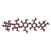
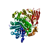
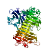
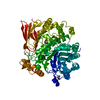
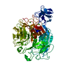
| Title | The X-ray crystal structure of a thermophilic cellobiose binding protein bound with cellopentaose | ||||||||||||
 Components Components | Oligopeptide ABC transporter, periplasmic oligopeptide-binding protein | ||||||||||||
 Keywords Keywords | SUGAR BINDING PROTEIN / cellulose / carbohydrate-binding protein / periplasmic binding protein / cellopentaose | ||||||||||||
| Function / homology |  Function and homology information Function and homology informationpeptide transport / peptide transmembrane transporter activity / metal ion binding Similarity search - Function | ||||||||||||
| Biological species |   Thermotoga maritima (bacteria) Thermotoga maritima (bacteria) | ||||||||||||
| Method |  X-RAY DIFFRACTION / X-RAY DIFFRACTION /  SYNCHROTRON / SYNCHROTRON /  MOLECULAR REPLACEMENT / Resolution: 1.5 Å MOLECULAR REPLACEMENT / Resolution: 1.5 Å | ||||||||||||
 Authors Authors | Cuneo, M.J. / Hellinga, H.W. | ||||||||||||
 Citation Citation |  Journal: J.Biol.Chem. / Year: 2009 Journal: J.Biol.Chem. / Year: 2009Title: Structural Analysis of Semi-specific Oligosaccharide Recognition by a Cellulose-binding Protein of Thermotoga maritima Reveals Adaptations for Functional Diversification of the Oligopeptide ...Title: Structural Analysis of Semi-specific Oligosaccharide Recognition by a Cellulose-binding Protein of Thermotoga maritima Reveals Adaptations for Functional Diversification of the Oligopeptide Periplasmic Binding Protein Fold. Authors: Cuneo, M.J. / Beese, L.S. / Hellinga, H.W. | ||||||||||||
| History |
|
- Structure visualization
| Structure viewer | Molecule:  Molmil Molmil Jmol/JSmol Jmol/JSmol |
|---|
- Downloads & links
- Download
| PDBx/mmCIF format |  3i5o.cif.gz 3i5o.cif.gz | 269.2 KB | Display |  PDBx/mmCIF format PDBx/mmCIF format |
|---|---|---|---|---|
| PDB format |  pdb3i5o.ent.gz pdb3i5o.ent.gz | 214.9 KB | Display |  PDB format PDB format |
| PDBx/mmJSON format |  3i5o.json.gz 3i5o.json.gz | Tree view |  PDBx/mmJSON format PDBx/mmJSON format | |
| Others |  Other downloads Other downloads |
-Validation report
| Summary document |  3i5o_validation.pdf.gz 3i5o_validation.pdf.gz | 942.2 KB | Display |  wwPDB validaton report wwPDB validaton report |
|---|---|---|---|---|
| Full document |  3i5o_full_validation.pdf.gz 3i5o_full_validation.pdf.gz | 952.3 KB | Display | |
| Data in XML |  3i5o_validation.xml.gz 3i5o_validation.xml.gz | 49.9 KB | Display | |
| Data in CIF |  3i5o_validation.cif.gz 3i5o_validation.cif.gz | 74.4 KB | Display | |
| Arichive directory |  https://data.pdbj.org/pub/pdb/validation_reports/i5/3i5o https://data.pdbj.org/pub/pdb/validation_reports/i5/3i5o ftp://data.pdbj.org/pub/pdb/validation_reports/i5/3i5o ftp://data.pdbj.org/pub/pdb/validation_reports/i5/3i5o | HTTPS FTP |
-Related structure data
| Related structure data | 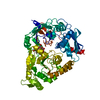 2o7iSC S: Starting model for refinement C: citing same article ( |
|---|---|
| Similar structure data |
- Links
Links
- Assembly
| Deposited unit | 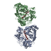
| |||||||||||||||||||||||
|---|---|---|---|---|---|---|---|---|---|---|---|---|---|---|---|---|---|---|---|---|---|---|---|---|
| 1 | 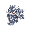
| |||||||||||||||||||||||
| 2 | 
| |||||||||||||||||||||||
| Unit cell |
| |||||||||||||||||||||||
| Noncrystallographic symmetry (NCS) | NCS domain:
NCS domain segments: Component-ID: 1 / Ens-ID: 1 / Beg auth comp-ID: SER / Beg label comp-ID: SER / End auth comp-ID: THR / End label comp-ID: THR / Auth seq-ID: 2 - 583 / Label seq-ID: 4 - 585
NCS oper: (Code: given Matrix: (-0.988763, -0.024936, -0.1474), Vector: Details | The biological unit is a single polypeptide chain. There are two biological units in the deposited files. | |
- Components
| #1: Protein | Mass: 68728.625 Da / Num. of mol.: 2 Source method: isolated from a genetically manipulated source Source: (gene. exp.)   Thermotoga maritima (bacteria) / Gene: tm0031, TM_0031 / Plasmid: pET21a / Production host: Thermotoga maritima (bacteria) / Gene: tm0031, TM_0031 / Plasmid: pET21a / Production host:  #2: Polysaccharide | #3: Water | ChemComp-HOH / | |
|---|
-Experimental details
-Experiment
| Experiment | Method:  X-RAY DIFFRACTION / Number of used crystals: 1 X-RAY DIFFRACTION / Number of used crystals: 1 |
|---|
- Sample preparation
| Crystal | Density Matthews: 2.48 Å3/Da / Density % sol: 50.32 % |
|---|---|
| Crystal grow | Temperature: 290 K / Method: vapor diffusion, hanging drop / pH: 6.5 Details: 0.1M NaCacodylate, pH 6.5, 0.2M Magnesium acetate, 20% PEG 4000, VAPOR DIFFUSION, HANGING DROP, temperature 290K |
-Data collection
| Diffraction | Mean temperature: 100 K |
|---|---|
| Diffraction source | Source:  SYNCHROTRON / Site: SYNCHROTRON / Site:  APS APS  / Beamline: 22-ID / Wavelength: 1 Å / Beamline: 22-ID / Wavelength: 1 Å |
| Detector | Type: MAR scanner 300 mm plate / Detector: IMAGE PLATE / Date: Dec 6, 2006 |
| Radiation | Monochromator: Si 111 / Protocol: SINGLE WAVELENGTH / Monochromatic (M) / Laue (L): M / Scattering type: x-ray |
| Radiation wavelength | Wavelength: 1 Å / Relative weight: 1 |
| Reflection | Resolution: 1.5→50 Å / Num. all: 205603 / Num. obs: 205603 / % possible obs: 96.3 % / Observed criterion σ(F): 0 / Observed criterion σ(I): -3 / Biso Wilson estimate: 22.456 Å2 / Rmerge(I) obs: 0.05 / Net I/σ(I): 15.7 |
| Reflection shell | Resolution: 1.5→1.6 Å / Rmerge(I) obs: 0.329 / Mean I/σ(I) obs: 4 / Num. measured obs: 93763 / Num. unique obs: 31437 / % possible all: 83.9 |
- Processing
| Software |
| |||||||||||||||||||||||||||||||||||||||||||||||||||||||||||||||||||||||||||||||||||||||||||||||||||||||||||||||||||||||||||||||||||||||||||||||||||||||||||||||||||||||||||||||||||||||||||||||||||||||||||||||||||||||||
|---|---|---|---|---|---|---|---|---|---|---|---|---|---|---|---|---|---|---|---|---|---|---|---|---|---|---|---|---|---|---|---|---|---|---|---|---|---|---|---|---|---|---|---|---|---|---|---|---|---|---|---|---|---|---|---|---|---|---|---|---|---|---|---|---|---|---|---|---|---|---|---|---|---|---|---|---|---|---|---|---|---|---|---|---|---|---|---|---|---|---|---|---|---|---|---|---|---|---|---|---|---|---|---|---|---|---|---|---|---|---|---|---|---|---|---|---|---|---|---|---|---|---|---|---|---|---|---|---|---|---|---|---|---|---|---|---|---|---|---|---|---|---|---|---|---|---|---|---|---|---|---|---|---|---|---|---|---|---|---|---|---|---|---|---|---|---|---|---|---|---|---|---|---|---|---|---|---|---|---|---|---|---|---|---|---|---|---|---|---|---|---|---|---|---|---|---|---|---|---|---|---|---|---|---|---|---|---|---|---|---|---|---|---|---|---|---|---|---|
| Refinement | Method to determine structure:  MOLECULAR REPLACEMENT MOLECULAR REPLACEMENTStarting model: 2O7I Resolution: 1.5→48.67 Å / Occupancy max: 1 / Occupancy min: 0.12 / FOM work R set: 0.802 / SU ML: 0.01 / σ(F): 1.99 / Stereochemistry target values: ML
| |||||||||||||||||||||||||||||||||||||||||||||||||||||||||||||||||||||||||||||||||||||||||||||||||||||||||||||||||||||||||||||||||||||||||||||||||||||||||||||||||||||||||||||||||||||||||||||||||||||||||||||||||||||||||
| Solvent computation | Shrinkage radii: 0.9 Å / VDW probe radii: 1.11 Å / Solvent model: FLAT BULK SOLVENT MODEL / Bsol: 49.652 Å2 / ksol: 0.367 e/Å3 | |||||||||||||||||||||||||||||||||||||||||||||||||||||||||||||||||||||||||||||||||||||||||||||||||||||||||||||||||||||||||||||||||||||||||||||||||||||||||||||||||||||||||||||||||||||||||||||||||||||||||||||||||||||||||
| Displacement parameters | Biso max: 71.44 Å2 / Biso mean: 23.436 Å2 / Biso min: 9 Å2
| |||||||||||||||||||||||||||||||||||||||||||||||||||||||||||||||||||||||||||||||||||||||||||||||||||||||||||||||||||||||||||||||||||||||||||||||||||||||||||||||||||||||||||||||||||||||||||||||||||||||||||||||||||||||||
| Refinement step | Cycle: LAST / Resolution: 1.5→48.67 Å
| |||||||||||||||||||||||||||||||||||||||||||||||||||||||||||||||||||||||||||||||||||||||||||||||||||||||||||||||||||||||||||||||||||||||||||||||||||||||||||||||||||||||||||||||||||||||||||||||||||||||||||||||||||||||||
| Refine LS restraints |
| |||||||||||||||||||||||||||||||||||||||||||||||||||||||||||||||||||||||||||||||||||||||||||||||||||||||||||||||||||||||||||||||||||||||||||||||||||||||||||||||||||||||||||||||||||||||||||||||||||||||||||||||||||||||||
| Refine LS restraints NCS |
| |||||||||||||||||||||||||||||||||||||||||||||||||||||||||||||||||||||||||||||||||||||||||||||||||||||||||||||||||||||||||||||||||||||||||||||||||||||||||||||||||||||||||||||||||||||||||||||||||||||||||||||||||||||||||
| LS refinement shell | Refine-ID: X-RAY DIFFRACTION / Total num. of bins used: 30
|
 Movie
Movie Controller





 PDBj
PDBj
