 Movie
Movie Controller

[English] 日本語
 Yorodumi
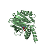
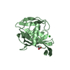
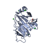
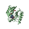
- PDB-3hyn: Crystal structure of a putative signal transduction protein (eubr... -
+ Open data

- Basic information
| Entry | Database: PDB / ID: 3hyn | ||||||
|---|---|---|---|---|---|---|---|
| Title | Crystal structure of a putative signal transduction protein (eubrec_0645) from eubacterium rectale atcc 33656 at 1.20 A resolution | ||||||
 Components Components | Putative signal transduction protein | ||||||
 Keywords Keywords | SIGNALING PROTEIN / Duf1863 family protein / nucleotide-binding protein / structural genomics / Joint Center for Structural Genomics / JCSG / Protein Structure Initiative / PSI-2 | ||||||
| Function / homology | Rossmann fold - #11200 / Thoeris protein ThsB, TIR-like domain / Thoeris protein ThsB, TIR-like domain / Rossmann fold / 3-Layer(aba) Sandwich / Alpha Beta / Thoeris protein ThsB TIR-like domain-containing protein Function and homology information Function and homology information | ||||||
| Biological species |  Eubacterium rectale ATCC 33656 (bacteria) Eubacterium rectale ATCC 33656 (bacteria) | ||||||
| Method |  X-RAY DIFFRACTION / SYNCHROTRON / SAD / Resolution: 1.2 Å X-RAY DIFFRACTION / SYNCHROTRON / SAD / Resolution: 1.2 Å | ||||||
 Authors Authors | Joint Center for Structural Genomics (JCSG) | ||||||
 Citation Citation | Journal: To be published Title: Crystal structure of Putative signal transduction protein (YP_002936568.1) from Eubacterium rectale at 1.20 A resolution Authors: Joint Center for Structural Genomics (JCSG) | ||||||
| History |
|
- Structure visualization
| Structure viewer | Molecule: MolmilJmol/JSmol |
|---|
- Downloads & links
-Download
| PDBx/mmCIF format | 3hyn.cif.gz | 110.1 KB | Display | PDBx/mmCIF format |
|---|---|---|---|---|
| PDB format | pdb3hyn.ent.gz | 84.6 KB | Display | PDB format |
| PDBx/mmJSON format | 3hyn.json.gz | Tree view | PDBx/mmJSON format | |
| Others |  Other downloads Other downloads |
-Validation report
| Arichive directory | https://data.pdbj.org/pub/pdb/validation_reports/hy/3hynftp://data.pdbj.org/pub/pdb/validation_reports/hy/3hyn | HTTPS FTP |
|---|
-Related structure data
| Similar structure data | |
|---|---|
| Other databases |


-Links
 PDBj
PDBj- Assembly
| Deposited unit | 
| ||||||||
|---|---|---|---|---|---|---|---|---|---|
| 1 |
| ||||||||
| Unit cell |
| ||||||||
| Details | ANALYTICAL SIZE EXCLUSION CHROMATOGRAPHY SUPPORTS THE ASSIGNMENT OF A MONOMER AS THE SIGNIFICANT OLIGOMERIZATION STATE IN SOLUTION. |
-Components
| #1: Protein | Mass: 22084.646 Da / Num. of mol.: 1 Source method: isolated from a genetically manipulated source Source: (gene. exp.) Eubacterium rectale ATCC 33656 (bacteria)Gene: YP_002936568.1 / Plasmid: SpeedET / Production host: |
|---|---|
| #2: Chemical | ChemComp-MRD / (  Mass: 118.174 Da / Num. of mol.: 1 / Source method: obtained synthetically / Formula: C6H14O2 / Comment: precipitant*YM Mass: 118.174 Da / Num. of mol.: 1 / Source method: obtained synthetically / Formula: C6H14O2 / Comment: precipitant*YM |
| #3: Water | ChemComp-HOH /  Mass: 18.015 Da / Num. of mol.: 301 / Source method: isolated from a natural source / Formula: H2O Mass: 18.015 Da / Num. of mol.: 301 / Source method: isolated from a natural source / Formula: H2O |
| Has protein modification | Y |
| Sequence details | THE CONSTRUCT WAS EXPRESSED WITH A PURIFICATION TAG MGSDKIHHHHHHENLYFQG. THE TAG WAS REMOVED WITH ...THE CONSTRUCT WAS EXPRESSED WITH A PURIFICATI |
-Experimental details
-Experiment
| Experiment | Method: X-RAY DIFFRACTION / Number of used crystals: 1 |
|---|
- Sample preparation
| Crystal | Density Matthews: 1.84 Å3/Da / Density % sol: 33.22 % |
|---|---|
| Crystal grow | Temperature: 277 K / Method: vapor diffusion, sitting drop / pH: 9 Details: 40.0000% MPD, 0.1M Bicine pH 9.0, NANODROP, VAPOR DIFFUSION, SITTING DROP, temperature 277K |
-Data collection
| Diffraction | Mean temperature: 100 K | ||||||||||||||||||||||||||||||||||||||||||||||||||||||||||||||||||
|---|---|---|---|---|---|---|---|---|---|---|---|---|---|---|---|---|---|---|---|---|---|---|---|---|---|---|---|---|---|---|---|---|---|---|---|---|---|---|---|---|---|---|---|---|---|---|---|---|---|---|---|---|---|---|---|---|---|---|---|---|---|---|---|---|---|---|---|
| Diffraction source | Source: SYNCHROTRON / Site: SSRL  / Beamline: BL9-2 / Wavelength: 0.97922 / Beamline: BL9-2 / Wavelength: 0.97922 | ||||||||||||||||||||||||||||||||||||||||||||||||||||||||||||||||||
| Detector | Type: MARMOSAIC 325 mm CCD / Detector: CCD / Date: May 13, 2009 / Details: Flat collimating mirror, toroid focusing mirror | ||||||||||||||||||||||||||||||||||||||||||||||||||||||||||||||||||
| Radiation | Monochromator: Double crystal monochromator / Protocol: SINGLE WAVELENGTH / Monochromatic (M) / Laue (L): M / Scattering type: x-ray | ||||||||||||||||||||||||||||||||||||||||||||||||||||||||||||||||||
| Radiation wavelength | Wavelength: 0.97922 Å / Relative weight: 1 | ||||||||||||||||||||||||||||||||||||||||||||||||||||||||||||||||||
| Reflection | Resolution: 1.2→32.461 Å / Num. obs: 51559 / % possible obs: 99.3 % / Observed criterion σ(I): -3 / Biso Wilson estimate: 8.178 Å2 / Rmerge(I) obs: 0.076 / Net I/σ(I): 9.1 | ||||||||||||||||||||||||||||||||||||||||||||||||||||||||||||||||||
| Reflection shell |
|
-Phasing
| Phasing | Method: SAD |
|---|
- Processing
| Software |
| ||||||||||||||||||||||||||||||||||||||||||||||||||||||||||||||||||||||||||||||||||||||||||
|---|---|---|---|---|---|---|---|---|---|---|---|---|---|---|---|---|---|---|---|---|---|---|---|---|---|---|---|---|---|---|---|---|---|---|---|---|---|---|---|---|---|---|---|---|---|---|---|---|---|---|---|---|---|---|---|---|---|---|---|---|---|---|---|---|---|---|---|---|---|---|---|---|---|---|---|---|---|---|---|---|---|---|---|---|---|---|---|---|---|---|---|
| Refinement | Method to determine structure: SAD / Resolution: 1.2→32.461 Å / Cor.coef. Fo:Fc: 0.982 / Cor.coef. Fo:Fc free: 0.978 / Occupancy max: 1 / Occupancy min: 0.17 / SU B: 1.059 / SU ML: 0.021 / Cross valid method: THROUGHOUT / σ(F): 0 / ESU R: 0.036 / ESU R Free: 0.034 Stereochemistry target values: MAXIMUM LIKELIHOOD WITH PHASES Details: 1. HYDROGENS HAVE BEEN ADDED IN THE RIDING POSITIONS. 2. A MET-INHIBITION PROTOCOL WAS USED FOR SELENOMETHIONINE INCORPORATION DURING PROTEIN EXPRESSION. THE OCCUPANCY OF THE SE ATOMS IN THE ...Details: 1. HYDROGENS HAVE BEEN ADDED IN THE RIDING POSITIONS. 2. A MET-INHIBITION PROTOCOL WAS USED FOR SELENOMETHIONINE INCORPORATION DURING PROTEIN EXPRESSION. THE OCCUPANCY OF THE SE ATOMS IN THE MSE RESIDUES WAS REDUCED TO 0.85 FOR THE REDUCED SCATTERING POWER DUE TO PARTIAL S-MET INCORPORATION. 3. (4R)-2-METHYLPENTANE-2,4-DIOL (MRD) MODELED IS PRESENT IN CRYSTALLIZATION SOLUTION. 4. CISPEPTIDE 28-29 ARE SUPPORTED BY DENSITY.
| ||||||||||||||||||||||||||||||||||||||||||||||||||||||||||||||||||||||||||||||||||||||||||
| Solvent computation | Ion probe radii: 0.8 Å / Shrinkage radii: 0.8 Å / VDW probe radii: 1.2 Å / Solvent model: MASK | ||||||||||||||||||||||||||||||||||||||||||||||||||||||||||||||||||||||||||||||||||||||||||
| Displacement parameters | Biso max: 77.73 Å2 / Biso mean: 12.96 Å2 / Biso min: 3.76 Å2
| ||||||||||||||||||||||||||||||||||||||||||||||||||||||||||||||||||||||||||||||||||||||||||
| Refinement step | Cycle: LAST / Resolution: 1.2→32.461 Å
| ||||||||||||||||||||||||||||||||||||||||||||||||||||||||||||||||||||||||||||||||||||||||||
| Refine LS restraints |
| ||||||||||||||||||||||||||||||||||||||||||||||||||||||||||||||||||||||||||||||||||||||||||
| LS refinement shell | Resolution: 1.2→1.231 Å / Total num. of bins used: 20
|
