 Movie
Movie Controller

[English] 日本語
 Yorodumi
- PDB-3hxq: Crystal Structure of Von Willebrand Factor (VWF) A1 Domain in Com... -
+ Open data

- Basic information
| Entry | Database: PDB / ID: 3hxq | ||||||
|---|---|---|---|---|---|---|---|
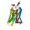
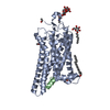
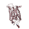
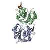
| Title | Crystal Structure of Von Willebrand Factor (VWF) A1 Domain in Complex with DNA Aptamer ARC1172, an Inhibitor of VWF-Platelet Binding | ||||||
 Components Components |
| ||||||
 Keywords Keywords | BLOOD CLOTTING/BLOOD CLOTTING REGULATOR / ARC1779 / VWF / Platelet Glycoprotein Ib / Aptamer / ARC1772 / Blood coagulation / Cell adhesion / Cleavage on pair of basic residues / Disease mutation / Disulfide bond / Extracellular matrix / Glycoprotein / Hemostasis / Isopeptide bond / Secreted / von Willebrand disease / BLOOD CLOTTING-BLOOD CLOTTING REGULATOR COMPLEX | ||||||
| Function / homology |  Function and homology information Function and homology informationDefective VWF binding to collagen type I / Enhanced cleavage of VWF variant by ADAMTS13 / Defective VWF cleavage by ADAMTS13 variant / Defective F8 binding to von Willebrand factor / Enhanced binding of GP1BA variant to VWF multimer:collagen / Defective binding of VWF variant to GPIb:IX:V / Weibel-Palade body / hemostasis / platelet alpha granule / Platelet Adhesion to exposed collagen ...Defective VWF binding to collagen type I / Enhanced cleavage of VWF variant by ADAMTS13 / Defective VWF cleavage by ADAMTS13 variant / Defective F8 binding to von Willebrand factor / Enhanced binding of GP1BA variant to VWF multimer:collagen / Defective binding of VWF variant to GPIb:IX:V / Weibel-Palade body / hemostasis / platelet alpha granule / Platelet Adhesion to exposed collagen / extracellular matrix structural constituent / GP1b-IX-V activation signalling / p130Cas linkage to MAPK signaling for integrins / Defective F8 cleavage by thrombin / Platelet Aggregation (Plug Formation) / cell-substrate adhesion / GRB2:SOS provides linkage to MAPK signaling for Integrins / positive regulation of intracellular signal transduction / immunoglobulin binding / Integrin cell surface interactions / collagen binding / : / Integrin signaling / platelet alpha granule lumen / Signaling by high-kinase activity BRAF mutants / platelet activation / MAP2K and MAPK activation / response to wounding / integrin binding / blood coagulation / Signaling by RAF1 mutants / Signaling by moderate kinase activity BRAF mutants / Paradoxical activation of RAF signaling by kinase inactive BRAF / Signaling downstream of RAS mutants / Signaling by BRAF and RAF1 fusions / Platelet degranulation / protein-folding chaperone binding / extracellular matrix / protease binding / cell adhesion / endoplasmic reticulum / : / extracellular exosome / extracellular region / identical protein binding Similarity search - Function | ||||||
| Biological species |  Homo sapiens (human) Homo sapiens (human) | ||||||
| Method |  X-RAY DIFFRACTION / MOLECULAR REPLACEMENT / Resolution: 2.694 Å X-RAY DIFFRACTION / MOLECULAR REPLACEMENT / Resolution: 2.694 Å | ||||||
 Authors Authors | Huang, R.H. / Sadler, J.E. / Fremont, D.H. / Diener, J.L. / Schaub, R.G. | ||||||
 Citation Citation | Journal: Structure / Year: 2009 Title: A structural explanation for the antithrombotic activity of ARC1172, a DNA aptamer that binds von Willebrand factor domain A1. Authors: Huang, R.H. / Fremont, D.H. / Diener, J.L. / Schaub, R.G. / Sadler, J.E. #1: Journal: Circulation / Year: 2007 Title: First-in-human evaluation of anti von Willebrand factor therapeutic aptamer ARC1779 in healthy volunteers. Authors: Gilbert, J.C. / DeFeo-Fraulini, T. / Hutabarat, R.M. / Horvath, C.J. / Merlino, P.G. / Marsh, H.N. / Healy, J.M. / Boufakhreddine, S. / Holohan, T.V. / Schaub, R.G. #2: Journal: J.Thromb.Haemost. / Year: 2009 Title: Inhibition of von Willebrand factor-mediated platelet activation and thrombosis by the anti-von Willebrand factor A1-domain aptamer ARC1779. Authors: Diener, J.L. / Daniel Lagasse, H.A. / Duerschmied, D. / Merhi, Y. / Tanguay, J.F. / Hutabarat, R. / Gilbert, J. / Wagner, D.D. / Schaub, R. | ||||||
| History |
|
- Structure visualization
| Structure viewer | Molecule: MolmilJmol/JSmol |
|---|
- Downloads & links
-Download
| PDBx/mmCIF format | 3hxq.cif.gz | 146.9 KB | Display | PDBx/mmCIF format |
|---|---|---|---|---|
| PDB format | pdb3hxq.ent.gz | 113.5 KB | Display | PDB format |
| PDBx/mmJSON format | 3hxq.json.gz | Tree view | PDBx/mmJSON format | |
| Others |  Other downloads Other downloads |
-Validation report
| Arichive directory | https://data.pdbj.org/pub/pdb/validation_reports/hx/3hxqftp://data.pdbj.org/pub/pdb/validation_reports/hx/3hxq | HTTPS FTP |
|---|
-Related structure data
| Related structure data |  3hxoSC S: Starting model for refinement C: citing same article ( |
|---|---|
| Similar structure data |

-Links
 PDBj
PDBj


- Assembly
| Deposited unit | 
| ||||||||
|---|---|---|---|---|---|---|---|---|---|
| 1 |
| ||||||||
| Unit cell |
| ||||||||
| Components on special symmetry positions |
|
-Components
| #1: Protein | Mass: 23889.627 Da / Num. of mol.: 1 Fragment: von Willebrand factor (VWF) A1 Domain (residues 1260-1468) Source method: isolated from a genetically manipulated source Source: (gene. exp.) Homo sapiens (human) / Production host:  |
|---|---|
| #2: DNA chain | Mass: 12884.188 Da / Num. of mol.: 1 / Fragment: Aptamer ARC1172 / Source method: obtained synthetically Details: ARC1172 is a DNA oligonucleotides that bind specific target molecules, identified by selection in vitro from large random sequence libraries (termed SELEX: systematic evolution of ligands by ...Details: ARC1172 is a DNA oligonucleotides that bind specific target molecules, identified by selection in vitro from large random sequence libraries (termed SELEX: systematic evolution of ligands by exponential enrichment) |
| #3: Water | ChemComp-HOH /  Mass: 18.015 Da / Num. of mol.: 102 / Source method: isolated from a natural source / Formula: H2O Mass: 18.015 Da / Num. of mol.: 102 / Source method: isolated from a natural source / Formula: H2O |
| Compound details | AUTHOR STATE THAT THE LAST RESIDUE OF THE DNA POLYMER IS AN INVERTED-DT WHICH IS NOT MODELED DUE TO ...AUTHOR STATE THAT THE LAST RESIDUE OF THE DNA POLYMER IS AN INVERTED-DT WHICH IS NOT MODELED DUE TO DISORDER. 3' INVERTED DT IS LINKED TO THE PHOSPHATE GROUP OF ITS PREVIOUS RESIDUE BY THE THIRD CARBON ATOMS OF THE SUGAR RINGS. |
| Has protein modification | Y |
-Experimental details
-Experiment
| Experiment | Method: X-RAY DIFFRACTION / Number of used crystals: 1 |
|---|
- Sample preparation
| Crystal | Density Matthews: 2.41 Å3/Da / Density % sol: 49.06 % |
|---|---|
| Crystal grow | Temperature: 293 K / pH: 4.6 Details: 0.2 M NH4Ac, 0.1 M NaAc, 30% PEG4000, pH 4.6, EVAPORATION, temperature 293K |
-Data collection
| Diffraction | Mean temperature: 100 K |
|---|---|
| Diffraction source | Source: ROTATING ANODE / Type: RIGAKU / Wavelength: 1.5418 |
| Detector | Type: RIGAKU RAXIS IV / Detector: IMAGE PLATE / Date: Mar 17, 2008 |
| Radiation | Monochromator: GRAPHITE / Protocol: SINGLE WAVELENGTH / Monochromatic (M) / Laue (L): M / Scattering type: x-ray |
| Radiation wavelength | Wavelength: 1.5418 Å / Relative weight: 1 |
| Reflection | Resolution: 2.694→38 Å / Num. obs: 9819 / % possible obs: 94.8 % / Observed criterion σ(I): 0 / Redundancy: 6.4 % / Rmerge(I) obs: 0.08 / Net I/σ(I): 17.2 |
| Reflection shell | Resolution: 2.69→2.8 Å / Redundancy: 4 % / Rmerge(I) obs: 0.316 / Mean I/σ(I) obs: 2.8 / % possible all: 83.9 |
- Processing
| Software |
| ||||||||||||||||||||||||||||||||||||||||||||||||||||||||||||||||||||||||||||||||||||||||||||||||||||
|---|---|---|---|---|---|---|---|---|---|---|---|---|---|---|---|---|---|---|---|---|---|---|---|---|---|---|---|---|---|---|---|---|---|---|---|---|---|---|---|---|---|---|---|---|---|---|---|---|---|---|---|---|---|---|---|---|---|---|---|---|---|---|---|---|---|---|---|---|---|---|---|---|---|---|---|---|---|---|---|---|---|---|---|---|---|---|---|---|---|---|---|---|---|---|---|---|---|---|---|---|---|
| Refinement | Method to determine structure: MOLECULAR REPLACEMENT Starting model: 3HXO Resolution: 2.694→38 Å / Occupancy max: 1 / Occupancy min: 0.47 / SU ML: 0.44 / σ(F): 1.5 / Phase error: 27.15 / Stereochemistry target values: ML
| ||||||||||||||||||||||||||||||||||||||||||||||||||||||||||||||||||||||||||||||||||||||||||||||||||||
| Solvent computation | Shrinkage radii: 0.9 Å / VDW probe radii: 1.11 Å / Solvent model: FLAT BULK SOLVENT MODEL / Bsol: 44.193 Å2 / ksol: 0.311 e/Å3 | ||||||||||||||||||||||||||||||||||||||||||||||||||||||||||||||||||||||||||||||||||||||||||||||||||||
| Displacement parameters | Biso max: 170.21 Å2 / Biso mean: 63.419 Å2 / Biso min: 17.48 Å2 | ||||||||||||||||||||||||||||||||||||||||||||||||||||||||||||||||||||||||||||||||||||||||||||||||||||
| Refinement step | Cycle: LAST / Resolution: 2.694→38 Å
| ||||||||||||||||||||||||||||||||||||||||||||||||||||||||||||||||||||||||||||||||||||||||||||||||||||
| Refine LS restraints |
| ||||||||||||||||||||||||||||||||||||||||||||||||||||||||||||||||||||||||||||||||||||||||||||||||||||
| LS refinement shell |
| ||||||||||||||||||||||||||||||||||||||||||||||||||||||||||||||||||||||||||||||||||||||||||||||||||||
| Refinement TLS params. | Method: refined / Refine-ID: X-RAY DIFFRACTION
| ||||||||||||||||||||||||||||||||||||||||||||||||||||||||||||||||||||||||||||||||||||||||||||||||||||
| Refinement TLS group |
|
