 Movie
Movie Controller


+ Open data

- Basic information
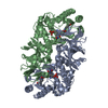
| Entry | Database: PDB / ID: 3hmk | ||||||
|---|---|---|---|---|---|---|---|
| Title | Crystal Structure of Serine Racemase | ||||||
 Components Components | Serine racemase | ||||||
 Keywords Keywords | ISOMERASE / Serine Racemase / D-serine / pyridoxal phosphate / PLP / NMDA glutamate receptor / schizophrenia | ||||||
| Function / homology |  Function and homology information Function and homology informationSerine metabolism / D-serine biosynthetic process / serine racemase / threonine racemase activity / response to ketamine / serine racemase activity / D-serine ammonia-lyase / L-serine ammonia-lyase / D-serine ammonia-lyase activity / L-serine ammonia-lyase activity ...Serine metabolism / D-serine biosynthetic process / serine racemase / threonine racemase activity / response to ketamine / serine racemase activity / D-serine ammonia-lyase / L-serine ammonia-lyase / D-serine ammonia-lyase activity / L-serine ammonia-lyase activity / : / pyruvate biosynthetic process / L-serine metabolic process / glycine binding / PDZ domain binding / apical part of cell / pyridoxal phosphate binding / response to lipopolysaccharide / response to xenobiotic stimulus / neuronal cell body / calcium ion binding / magnesium ion binding / protein homodimerization activity / ATP binding / identical protein binding / cytoplasm Similarity search - Function | ||||||
| Biological species |  | ||||||
| Method |  X-RAY DIFFRACTION / SYNCHROTRON / MOLECULAR REPLACEMENT / Resolution: 2.1 Å X-RAY DIFFRACTION / SYNCHROTRON / MOLECULAR REPLACEMENT / Resolution: 2.1 Å | ||||||
 Authors Authors | Smith, M.A. / Barker, J. / Mack, V. / Ebneth, A. / Felicetti, B. / Woods, M. | ||||||
 Citation Citation | Journal: J.Biol.Chem. / Year: 2010 Title: The structure of mammalian serine racemase: evidence for conformational changes upon inhibitor binding. Authors: Smith, M.A. / Mack, V. / Ebneth, A. / Moraes, I. / Felicetti, B. / Wood, M. / Schonfeld, D. / Mather, O. / Cesura, A. / Barker, J. | ||||||
| History |
|
- Structure visualization
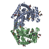
| Structure viewer | Molecule: MolmilJmol/JSmol |
|---|
- Downloads & links
-Download
| PDBx/mmCIF format | 3hmk.cif.gz | 136.7 KB | Display | PDBx/mmCIF format |
|---|---|---|---|---|
| PDB format | pdb3hmk.ent.gz | 105.5 KB | Display | PDB format |
| PDBx/mmJSON format | 3hmk.json.gz | Tree view | PDBx/mmJSON format | |
| Others |  Other downloads Other downloads |
-Validation report
| Arichive directory | https://data.pdbj.org/pub/pdb/validation_reports/hm/3hmkftp://data.pdbj.org/pub/pdb/validation_reports/hm/3hmk | HTTPS FTP |
|---|
-Related structure data
| Related structure data |  3l6bC  3l6cC  3l6rC 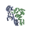 1fkpS  3fda 3fkp S: Starting model for refinement C: citing same article ( |
|---|---|
| Similar structure data |


-Links
 PDBj
PDBj
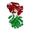
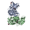
- Assembly
| Deposited unit | 
| ||||||||
|---|---|---|---|---|---|---|---|---|---|
| 1 |
| ||||||||
| Unit cell |
|
-Components
| #1: Protein | Mass: 36579.703 Da / Num. of mol.: 2 / Mutation: C2D, C6D Source method: isolated from a genetically manipulated source Source: (gene. exp.)  #2: Chemical |   Mass: 54.938 Da / Num. of mol.: 2 / Source method: obtained synthetically / Formula: Mn Mass: 54.938 Da / Num. of mol.: 2 / Source method: obtained synthetically / Formula: Mn#3: Chemical |   Mass: 247.142 Da / Num. of mol.: 2 / Source method: obtained synthetically / Formula: C8H10NO6P Mass: 247.142 Da / Num. of mol.: 2 / Source method: obtained synthetically / Formula: C8H10NO6P#4: Water | ChemComp-HOH / |  Mass: 18.015 Da / Num. of mol.: 202 / Source method: isolated from a natural source / Formula: H2O Mass: 18.015 Da / Num. of mol.: 202 / Source method: isolated from a natural source / Formula: H2O |
|---|
-Experimental details
-Experiment
| Experiment | Method: X-RAY DIFFRACTION / Number of used crystals: 1 |
|---|
- Sample preparation
| Crystal | Density Matthews: 2.05 Å3/Da / Density % sol: 40.09 % |
|---|---|
| Crystal grow | Temperature: 293 K / Method: vapor diffusion, hanging drop / pH: 8 Details: pH 8.0, VAPOR DIFFUSION, HANGING DROP, temperature 293K |
-Data collection
| Diffraction | Mean temperature: 100 K |
|---|---|
| Diffraction source | Source: SYNCHROTRON / Site: Diamond  / Beamline: I03 / Wavelength: 1.0332 Å / Beamline: I03 / Wavelength: 1.0332 Å |
| Detector | Type: ADSC QUANTUM 315 / Detector: CCD / Date: Sep 17, 2008 |
| Radiation | Monochromator: Double Crystal Silicon / Protocol: SINGLE WAVELENGTH / Monochromatic (M) / Laue (L): M / Scattering type: x-ray |
| Radiation wavelength | Wavelength: 1.0332 Å / Relative weight: 1 |
| Reflection | Resolution: 2.1→41.1 Å / Num. all: 34466 / Num. obs: 32657 / % possible obs: 95.8 % / Observed criterion σ(F): 2 / Observed criterion σ(I): 2 / Redundancy: 3.17 % / Biso Wilson estimate: 23.8 Å2 / Rmerge(I) obs: 0.069 / Rsym value: 0.069 / Net I/σ(I): 10.1 |
| Reflection shell | Resolution: 2.1→2.18 Å / Redundancy: 1.95 % / Rmerge(I) obs: 0.195 / Mean I/σ(I) obs: 3.5 / Num. unique all: 1837 / Rsym value: 0.195 / % possible all: 74.8 |
- Processing
| Software |
| |||||||||||||||||||||||||||||||||||||||||||||||||||||||||||||||||
|---|---|---|---|---|---|---|---|---|---|---|---|---|---|---|---|---|---|---|---|---|---|---|---|---|---|---|---|---|---|---|---|---|---|---|---|---|---|---|---|---|---|---|---|---|---|---|---|---|---|---|---|---|---|---|---|---|---|---|---|---|---|---|---|---|---|---|
| Refinement | Method to determine structure: MOLECULAR REPLACEMENT Starting model: PDB ENTRY 1FKP Resolution: 2.1→41.1 Å / Cor.coef. Fo:Fc: 0.92 / Cor.coef. Fo:Fc free: 0.881 / SU B: 6.637 / SU ML: 0.176 / Cross valid method: THROUGHOUT / σ(F): 2 / σ(I): 2 / ESU R: 0.336 / ESU R Free: 0.248 / Stereochemistry target values: MAXIMUM LIKELIHOOD / Details: HYDROGENS HAVE BEEN ADDED IN THE RIDING POSITIONS
| |||||||||||||||||||||||||||||||||||||||||||||||||||||||||||||||||
| Solvent computation | Ion probe radii: 0.8 Å / Shrinkage radii: 0.8 Å / VDW probe radii: 1.2 Å / Solvent model: MASK | |||||||||||||||||||||||||||||||||||||||||||||||||||||||||||||||||
| Displacement parameters | Biso mean: 29.478 Å2
| |||||||||||||||||||||||||||||||||||||||||||||||||||||||||||||||||
| Refine analyze | Luzzati coordinate error obs: 0.314 Å | |||||||||||||||||||||||||||||||||||||||||||||||||||||||||||||||||
| Refinement step | Cycle: LAST / Resolution: 2.1→41.1 Å
| |||||||||||||||||||||||||||||||||||||||||||||||||||||||||||||||||
| Refine LS restraints |
| |||||||||||||||||||||||||||||||||||||||||||||||||||||||||||||||||
| LS refinement shell | Resolution: 2.1→2.154 Å / Total num. of bins used: 20
|
