 Movie
Movie Controller

[English] 日本語
 Yorodumi
- PDB-3hji: 1.8 Angstrom Crystal Structure of the I74V:I85V Variant of Vivid ... -
+ Open data

- Basic information
| Entry | Database: PDB / ID: 3hji | ||||||
|---|---|---|---|---|---|---|---|
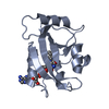
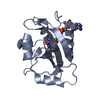
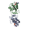
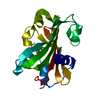
| Title | 1.8 Angstrom Crystal Structure of the I74V:I85V Variant of Vivid (VVD). | ||||||
 Components Components | Vivid PAS protein VVD | ||||||
 Keywords Keywords | SIGNALING PROTEIN / Photoreceptor / Circadian Clock / LOV / FAD | ||||||
| Function / homology |  Function and homology information Function and homology information | ||||||
| Biological species |  Neurospora crassa (fungus) Neurospora crassa (fungus) | ||||||
| Method |  X-RAY DIFFRACTION / SYNCHROTRON / MOLECULAR REPLACEMENT / Resolution: 1.8 Å X-RAY DIFFRACTION / SYNCHROTRON / MOLECULAR REPLACEMENT / Resolution: 1.8 Å | ||||||
 Authors Authors | Zoltowski, B.D. / Vaccaro, B.J. / Crane, B.R. | ||||||
 Citation Citation | Journal: Nat.Chem.Biol. / Year: 2009 Title: Mechanism-based tuning of a LOV domain photoreceptor. Authors: Zoltowski, B.D. / Vaccaro, B. / Crane, B.R. | ||||||
| History |
|
- Structure visualization
| Structure viewer | Molecule: MolmilJmol/JSmol |
|---|
- Downloads & links
-Download
| PDBx/mmCIF format | 3hji.cif.gz | 82 KB | Display | PDBx/mmCIF format |
|---|---|---|---|---|
| PDB format | pdb3hji.ent.gz | 60.3 KB | Display | PDB format |
| PDBx/mmJSON format | 3hji.json.gz | Tree view | PDBx/mmJSON format | |
| Others |  Other downloads Other downloads |
-Validation report
| Arichive directory | https://data.pdbj.org/pub/pdb/validation_reports/hj/3hjiftp://data.pdbj.org/pub/pdb/validation_reports/hj/3hji | HTTPS FTP |
|---|
-Related structure data
| Related structure data |  3hjkC 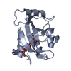 2pd7S C: citing same article ( S: Starting model for refinement |
|---|---|
| Similar structure data |


-Links
 PDBj
PDBj

- Assembly
| Deposited unit | 
| ||||||||
|---|---|---|---|---|---|---|---|---|---|
| 1 | 
| ||||||||
| 2 | 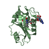
| ||||||||
| 3 |
| ||||||||
| Unit cell |
| ||||||||
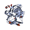
| Details | The protein in monomeric, however following photo-excitation the protein dimerizes. The biological dimer structure is currently unknown. |
-Components
| #1: Protein | Mass: 17484.949 Da / Num. of mol.: 2 / Mutation: I74V, I85V Source method: isolated from a genetically manipulated source Source: (gene. exp.) Neurospora crassa (fungus) / Gene: G17A4.050, Vivid, vvd / Plasmid: pET28 / Production host:  #2: Chemical |   Mass: 785.550 Da / Num. of mol.: 2 / Source method: obtained synthetically / Formula: C27H33N9O15P2 / Comment: FAD*YM Mass: 785.550 Da / Num. of mol.: 2 / Source method: obtained synthetically / Formula: C27H33N9O15P2 / Comment: FAD*YM#3: Water | ChemComp-HOH / |  Mass: 18.015 Da / Num. of mol.: 349 / Source method: isolated from a natural source / Formula: H2O Mass: 18.015 Da / Num. of mol.: 349 / Source method: isolated from a natural source / Formula: H2O |
|---|
-Experimental details
-Experiment
| Experiment | Method: X-RAY DIFFRACTION / Number of used crystals: 1 |
|---|
- Sample preparation
| Crystal | Density Matthews: 2.47 Å3/Da / Density % sol: 50.16 % |
|---|---|
| Crystal grow | Temperature: 298 K / Method: vapor diffusion, hanging drop / pH: 5.6 Details: 3.6 mg/ml protein in buffer containing 5 mM DTT, 100 mM NaCl, 50 mM Hepes pH 8.0 and 10% glycerol combined with equal volume of 28% PEG 4k, 100 mM ammonium acetate, and 100 mM tri-sodium ...Details: 3.6 mg/ml protein in buffer containing 5 mM DTT, 100 mM NaCl, 50 mM Hepes pH 8.0 and 10% glycerol combined with equal volume of 28% PEG 4k, 100 mM ammonium acetate, and 100 mM tri-sodium citrate pH 5.6, VAPOR DIFFUSION, HANGING DROP, temperature 298K |
-Data collection
| Diffraction | Mean temperature: 80 K |
|---|---|
| Diffraction source | Source: SYNCHROTRON / Site: CHESS  / Beamline: F2 / Wavelength: 0.979 Å / Beamline: F2 / Wavelength: 0.979 Å |
| Detector | Type: ADSC QUANTUM 210 / Detector: CCD / Date: May 1, 2008 |
| Radiation | Monochromator: Double-bounce downward, offset 25.4 mm / Protocol: SINGLE WAVELENGTH / Monochromatic (M) / Laue (L): M / Scattering type: x-ray |
| Radiation wavelength | Wavelength: 0.979 Å / Relative weight: 1 |
| Reflection | Resolution: 1.76→46.6 Å / Num. all: 29885 / Num. obs: 29885 / % possible obs: 97.8 % / Observed criterion σ(F): 0 / Observed criterion σ(I): 0 / Redundancy: 4.3 % / Biso Wilson estimate: 29.9 Å2 / Rmerge(I) obs: 0.063 / Net I/σ(I): 31 |
| Reflection shell | Resolution: 1.76→1.83 Å / Redundancy: 3.7 % / Rmerge(I) obs: 0.474 / Mean I/σ(I) obs: 2.5 / Num. unique all: 3502 / % possible all: 96.6 |
- Processing
| Software |
| |||||||||||||||||||||||||
|---|---|---|---|---|---|---|---|---|---|---|---|---|---|---|---|---|---|---|---|---|---|---|---|---|---|---|
| Refinement | Method to determine structure: MOLECULAR REPLACEMENT Starting model: 2pd7 Resolution: 1.8→30 Å / σ(F): 0 / Stereochemistry target values: Engh & Huber
| |||||||||||||||||||||||||
| Displacement parameters | Biso mean: 29.9 Å2 | |||||||||||||||||||||||||
| Refinement step | Cycle: LAST / Resolution: 1.8→30 Å
|
