 Movie
Movie Controller

[English] 日本語
 Yorodumi
- PDB-3hiw: Crystal structure of Saporin-L1 in complex with the cyclic tetran... -
+ Open data

- Basic information
| Entry | Database: PDB / ID: 3hiw | ||||||
|---|---|---|---|---|---|---|---|
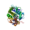
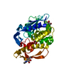
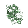
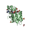
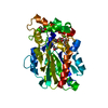
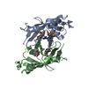
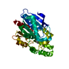
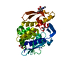
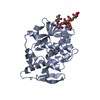
| Title | Crystal structure of Saporin-L1 in complex with the cyclic tetranucleotide inhibitor, a transition state analogue | ||||||
 Components Components | Vacuolar saporin | ||||||
 Keywords Keywords | Hydrolase/Hydrolase inhibitor / transition state / ribosome inactivating proteins / RIPs / Hydrolase / Plant defense / Protein synthesis inhibitor / Toxin / Hydrolase-Hydrolase inhibitor COMPLEX | ||||||
| Function / homology |  Function and homology information Function and homology informationrRNA N-glycosylase / rRNA N-glycosylase activity / defense response / toxin activity / negative regulation of translation Similarity search - Function | ||||||
| Biological species |  Saponaria officinalis (common soapwort) Saponaria officinalis (common soapwort) | ||||||
| Method |  X-RAY DIFFRACTION / SYNCHROTRON / MOLECULAR REPLACEMENT / Resolution: 1.8 Å X-RAY DIFFRACTION / SYNCHROTRON / MOLECULAR REPLACEMENT / Resolution: 1.8 Å | ||||||
 Authors Authors | Ho, M. / Sturm, M.B. / Almo, S.C. / Schramm, V.L. | ||||||
 Citation Citation | Journal: Proc.Natl.Acad.Sci.USA / Year: 2009 Title: Transition state analogues in structures of ricin and saporin ribosome-inactivating proteins. Authors: Ho, M.C. / Sturm, M.B. / Almo, S.C. / Schramm, V.L. | ||||||
| History |
|
- Structure visualization
| Structure viewer | Molecule: MolmilJmol/JSmol |
|---|
- Downloads & links
-Download
| PDBx/mmCIF format | 3hiw.cif.gz | 126.4 KB | Display | PDBx/mmCIF format |
|---|---|---|---|---|
| PDB format | pdb3hiw.ent.gz | 99.1 KB | Display | PDB format |
| PDBx/mmJSON format | 3hiw.json.gz | Tree view | PDBx/mmJSON format | |
| Others |  Other downloads Other downloads |
-Validation report
| Arichive directory | https://data.pdbj.org/pub/pdb/validation_reports/hi/3hiwftp://data.pdbj.org/pub/pdb/validation_reports/hi/3hiw | HTTPS FTP |
|---|
-Related structure data


-Links
 PDBj
PDBj

- Assembly
| Deposited unit | 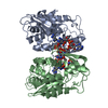
| ||||||||
|---|---|---|---|---|---|---|---|---|---|
| 1 | 
| ||||||||
| 2 | 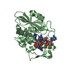
| ||||||||
| Unit cell |
|
-Components
| #1: Protein | Mass: 28767.773 Da / Num. of mol.: 2 Source method: isolated from a genetically manipulated source Source: (gene. exp.) Saponaria officinalis (common soapwort)Production host:  #2: Chemical |   Mass: 1598.109 Da / Num. of mol.: 2 / Source method: obtained synthetically / Formula: C51H72N21O29P5 Mass: 1598.109 Da / Num. of mol.: 2 / Source method: obtained synthetically / Formula: C51H72N21O29P5#3: Water | ChemComp-HOH / |  Mass: 18.015 Da / Num. of mol.: 442 / Source method: isolated from a natural source / Formula: H2O Mass: 18.015 Da / Num. of mol.: 442 / Source method: isolated from a natural source / Formula: H2O |
|---|
-Experimental details
-Experiment
| Experiment | Method: X-RAY DIFFRACTION / Number of used crystals: 1 |
|---|
- Sample preparation
| Crystal | Density Matthews: 2.25 Å3/Da / Density % sol: 45.25 % |
|---|---|
| Crystal grow | Temperature: 291 K / Method: vapor diffusion, sitting drop / pH: 5.5 Details: 20% PEG2000MME, 0.1M sodium acetate, 0.4M potassium thiocyanate, pH 5.5, VAPOR DIFFUSION, SITTING DROP, temperature 291K |
-Data collection
| Diffraction | Mean temperature: 100 K | ||||||||||||||||||||||||||||||||||||||||||||||||||||||||||||||||||
|---|---|---|---|---|---|---|---|---|---|---|---|---|---|---|---|---|---|---|---|---|---|---|---|---|---|---|---|---|---|---|---|---|---|---|---|---|---|---|---|---|---|---|---|---|---|---|---|---|---|---|---|---|---|---|---|---|---|---|---|---|---|---|---|---|---|---|---|
| Diffraction source | Source: SYNCHROTRON / Site: NSLS  / Beamline: X29A / Wavelength: 0.979 Å / Beamline: X29A / Wavelength: 0.979 Å | ||||||||||||||||||||||||||||||||||||||||||||||||||||||||||||||||||
| Detector | Type: ADSC QUANTUM 315 / Detector: CCD / Date: Oct 2, 2008 | ||||||||||||||||||||||||||||||||||||||||||||||||||||||||||||||||||
| Radiation | Protocol: SINGLE WAVELENGTH / Monochromatic (M) / Laue (L): M / Scattering type: x-ray | ||||||||||||||||||||||||||||||||||||||||||||||||||||||||||||||||||
| Radiation wavelength | Wavelength: 0.979 Å / Relative weight: 1 | ||||||||||||||||||||||||||||||||||||||||||||||||||||||||||||||||||
| Reflection | Resolution: 1.8→20 Å / Num. obs: 44853 / % possible obs: 97.1 % / Redundancy: 1.9 % / Rmerge(I) obs: 0.041 / Χ2: 1.031 / Net I/σ(I): 18.983 | ||||||||||||||||||||||||||||||||||||||||||||||||||||||||||||||||||
| Reflection shell |
|
- Processing
| Software |
| |||||||||||||||||||||||||||||||||||||||||||||||||||||||||||||||||
|---|---|---|---|---|---|---|---|---|---|---|---|---|---|---|---|---|---|---|---|---|---|---|---|---|---|---|---|---|---|---|---|---|---|---|---|---|---|---|---|---|---|---|---|---|---|---|---|---|---|---|---|---|---|---|---|---|---|---|---|---|---|---|---|---|---|---|
| Refinement | Method to determine structure: MOLECULAR REPLACEMENT / Resolution: 1.8→19.64 Å / Cor.coef. Fo:Fc: 0.945 / Cor.coef. Fo:Fc free: 0.918 / Occupancy max: 1 / Occupancy min: 0.5 / SU B: 2.65 / SU ML: 0.084 / Cross valid method: THROUGHOUT / σ(F): 0 / ESU R: 0.15 / ESU R Free: 0.138 / Stereochemistry target values: MAXIMUM LIKELIHOOD / Details: HYDROGENS HAVE BEEN ADDED IN THE RIDING POSITIONS
| |||||||||||||||||||||||||||||||||||||||||||||||||||||||||||||||||
| Solvent computation | Ion probe radii: 0.8 Å / Shrinkage radii: 0.8 Å / VDW probe radii: 1.2 Å / Solvent model: MASK | |||||||||||||||||||||||||||||||||||||||||||||||||||||||||||||||||
| Displacement parameters | Biso max: 63.23 Å2 / Biso mean: 17.906 Å2 / Biso min: 7.02 Å2
| |||||||||||||||||||||||||||||||||||||||||||||||||||||||||||||||||
| Refinement step | Cycle: LAST / Resolution: 1.8→19.64 Å
| |||||||||||||||||||||||||||||||||||||||||||||||||||||||||||||||||
| Refine LS restraints |
| |||||||||||||||||||||||||||||||||||||||||||||||||||||||||||||||||
| LS refinement shell | Resolution: 1.8→1.847 Å / Total num. of bins used: 20
|
