 Movie
Movie Controller

[English] 日本語
 Yorodumi
- PDB-3h8y: Crystal structure of carboxysome small shell protein CsoS1C from ... -
+ Open data

- Basic information
| Entry | Database: PDB / ID: 3h8y | ||||||
|---|---|---|---|---|---|---|---|
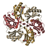
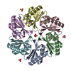
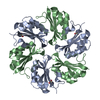
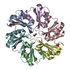
| Title | Crystal structure of carboxysome small shell protein CsoS1C from Halothiobacillus neapolitanus | ||||||
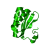
 Components Components | Major carboxysome shell protein 1C | ||||||
 Keywords Keywords | STRUCTURAL PROTEIN / bacterial microcompartment domain | ||||||
| Function / homology |  Function and homology information Function and homology information | ||||||
| Biological species |  Halothiobacillus neapolitanus (bacteria) Halothiobacillus neapolitanus (bacteria) | ||||||
| Method |  X-RAY DIFFRACTION / SYNCHROTRON / MOLECULAR REPLACEMENT / molecular replacement / Resolution: 2.51 Å X-RAY DIFFRACTION / SYNCHROTRON / MOLECULAR REPLACEMENT / molecular replacement / Resolution: 2.51 Å | ||||||
 Authors Authors | Tsai, Y. / Sawaya, M.R. / Yeates, T.O. | ||||||
 Citation Citation | Journal: Acta Crystallogr.,Sect.D / Year: 2009 Title: Analysis of lattice-translocation disorder in the layered hexagonal structure of carboxysome shell protein CsoS1C Authors: Tsai, Y. / Sawaya, M.R. / Yeates, T.O. | ||||||
| History |
|
- Structure visualization
| Structure viewer | Molecule: MolmilJmol/JSmol |
|---|
- Downloads & links
-Download
| PDBx/mmCIF format | 3h8y.cif.gz | 29.6 KB | Display | PDBx/mmCIF format |
|---|---|---|---|---|
| PDB format | pdb3h8y.ent.gz | 19.1 KB | Display | PDB format |
| PDBx/mmJSON format | 3h8y.json.gz | Tree view | PDBx/mmJSON format | |
| Others |  Other downloads Other downloads |
-Validation report
| Arichive directory | https://data.pdbj.org/pub/pdb/validation_reports/h8/3h8yftp://data.pdbj.org/pub/pdb/validation_reports/h8/3h8y | HTTPS FTP |
|---|
-Related structure data
| Related structure data |  2g13S S: Starting model for refinement |
|---|---|
| Similar structure data |


-Links
 PDBj
PDBj

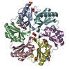
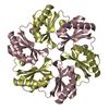
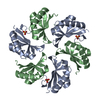
- Assembly
| Deposited unit | 
| ||||||||
|---|---|---|---|---|---|---|---|---|---|
| 1 | x 6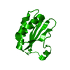
| ||||||||
| Unit cell |
| ||||||||
| Components on special symmetry positions |
|
-Components
| #1: Protein | Mass: 9930.453 Da / Num. of mol.: 1 Source method: isolated from a genetically manipulated source Source: (gene. exp.) Halothiobacillus neapolitanus (bacteria)Gene: csoS1C / Plasmid: pPROEx-Htb / Production host: |
|---|---|
| #2: Water | ChemComp-HOH /  Mass: 18.015 Da / Num. of mol.: 24 / Source method: isolated from a natural source / Formula: H2O Mass: 18.015 Da / Num. of mol.: 24 / Source method: isolated from a natural source / Formula: H2O |
-Experimental details
-Experiment
| Experiment | Method: X-RAY DIFFRACTION / Number of used crystals: 1 |
|---|
- Sample preparation
| Crystal | Density Matthews: 4.07 Å3/Da / Density % sol: 69.79 % |
|---|---|
| Crystal grow | Temperature: 298 K / Method: vapor diffusion, hanging drop / pH: 8.5 Details: 0.1M Tris HCl pH 8.5, 0.15M Sodium Citrate, 25% PEG 400, 0.01M nickel (II) chloride, vapor diffusion, hanging drop, temperature 298K |
-Data collection
| Diffraction | Mean temperature: 100 K |
|---|---|
| Diffraction source | Source: SYNCHROTRON / Site: APS  / Beamline: 24-ID-C / Wavelength: 0.9792 Å / Beamline: 24-ID-C / Wavelength: 0.9792 Å |
| Detector | Type: ADSC QUANTUM 315 / Detector: CCD / Date: Feb 25, 2008 |
| Radiation | Protocol: SINGLE WAVELENGTH / Monochromatic (M) / Laue (L): M / Scattering type: x-ray |
| Radiation wavelength | Wavelength: 0.9792 Å / Relative weight: 1 |
| Reflection | Resolution: 2.5→90 Å / Num. all: 5538 / Num. obs: 5538 / % possible obs: 100 % / Observed criterion σ(I): -3 / Redundancy: 8.3 % / Biso Wilson estimate: 37.6 Å2 / Rmerge(I) obs: 0.186 / Χ2: 1.272 / Net I/σ(I): 13.17 |
| Reflection shell | Resolution: 2.5→2.69 Å / Redundancy: 8.4 % / Rmerge(I) obs: 0.359 / Mean I/σ(I) obs: 6.94 / Num. unique all: 1103 / Χ2: 1.18 / % possible all: 100 |
-Phasing
| Phasing | Method: molecular replacement | |||||||||
|---|---|---|---|---|---|---|---|---|---|---|
| Phasing MR | Model details: Phaser MODE: MR_AUTO
|
- Processing
| Software |
| |||||||||||||||||||||||||||||||||||||||||||||||||||||||||||||||||||||||||||||||||||||
|---|---|---|---|---|---|---|---|---|---|---|---|---|---|---|---|---|---|---|---|---|---|---|---|---|---|---|---|---|---|---|---|---|---|---|---|---|---|---|---|---|---|---|---|---|---|---|---|---|---|---|---|---|---|---|---|---|---|---|---|---|---|---|---|---|---|---|---|---|---|---|---|---|---|---|---|---|---|---|---|---|---|---|---|---|---|---|
| Refinement | Method to determine structure: MOLECULAR REPLACEMENT Starting model: PDB entry 2G13 Resolution: 2.51→26.24 Å / Cor.coef. Fo:Fc: 0.909 / Cor.coef. Fo:Fc free: 0.833 / WRfactor Rfree: 0.322 / WRfactor Rwork: 0.274 / Occupancy max: 1 / Occupancy min: 0.17 / FOM work R set: 0.554 / SU B: 14.163 / SU ML: 0.342 / SU R Cruickshank DPI: 0.37 / SU Rfree: 0.301 / Cross valid method: THROUGHOUT / σ(F): 0 / ESU R: 0.37 / ESU R Free: 0.3 / Stereochemistry target values: MAXIMUM LIKELIHOOD / Details: HYDROGENS HAVE BEEN ADDED IN THE RIDING POSITIONS
| |||||||||||||||||||||||||||||||||||||||||||||||||||||||||||||||||||||||||||||||||||||
| Solvent computation | Ion probe radii: 0.8 Å / Shrinkage radii: 0.8 Å / VDW probe radii: 1.4 Å / Solvent model: MASK | |||||||||||||||||||||||||||||||||||||||||||||||||||||||||||||||||||||||||||||||||||||
| Displacement parameters | Biso max: 79.65 Å2 / Biso mean: 35.061 Å2 / Biso min: 2 Å2
| |||||||||||||||||||||||||||||||||||||||||||||||||||||||||||||||||||||||||||||||||||||
| Refinement step | Cycle: LAST / Resolution: 2.51→26.24 Å
| |||||||||||||||||||||||||||||||||||||||||||||||||||||||||||||||||||||||||||||||||||||
| Refine LS restraints |
| |||||||||||||||||||||||||||||||||||||||||||||||||||||||||||||||||||||||||||||||||||||
| LS refinement shell | Resolution: 2.509→2.574 Å / Total num. of bins used: 20
|
