 Movie
Movie Controller


+ Open data

- Basic information
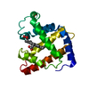
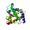
| Entry | Database: PDB / ID: 3h58 | ||||||
|---|---|---|---|---|---|---|---|
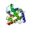
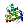
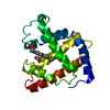
| Title | Myoglobin Cavity Mutant H64LV68N Met form | ||||||
 Components Components | Myoglobin | ||||||
 Keywords Keywords | OXYGEN STORAGE / OXYGEN TRANSPORT / Myoglobin / active site hydration / ligand entry and exit / oxygen storage and transport / Heme / Iron / Metal-binding / Muscle protein / Transport | ||||||
| Function / homology |  Function and homology information Function and homology informationOxidoreductases; Acting on other nitrogenous compounds as donors / nitrite reductase activity / sarcoplasm / Oxidoreductases; Acting on a peroxide as acceptor; Peroxidases / removal of superoxide radicals / oxygen carrier activity / peroxidase activity / oxygen binding / heme binding / extracellular exosome / metal ion binding Similarity search - Function | ||||||
| Biological species |  | ||||||
| Method |  X-RAY DIFFRACTION / FOURIER SYNTHESIS / Resolution: 1.8 Å X-RAY DIFFRACTION / FOURIER SYNTHESIS / Resolution: 1.8 Å | ||||||
 Authors Authors | Soman, J. / Olson, J.S. | ||||||
 Citation Citation | Journal: J.Am.Chem.Soc. / Year: 2009 Title: Optical detection of disordered water within a protein cavity. Authors: Goldbeck, R.A. / Pillsbury, M.L. / Jensen, R.A. / Mendoza, J.L. / Nguyen, R.L. / Olson, J.S. / Soman, J. / Kliger, D.S. / Esquerra, R.M. | ||||||
| History |
|
- Structure visualization
| Structure viewer | Molecule: MolmilJmol/JSmol |
|---|
- Downloads & links
-Download
| PDBx/mmCIF format | 3h58.cif.gz | 52.6 KB | Display | PDBx/mmCIF format |
|---|---|---|---|---|
| PDB format | pdb3h58.ent.gz | 35.6 KB | Display | PDB format |
| PDBx/mmJSON format | 3h58.json.gz | Tree view | PDBx/mmJSON format | |
| Others |  Other downloads Other downloads |
-Validation report
| Arichive directory | https://data.pdbj.org/pub/pdb/validation_reports/h5/3h58ftp://data.pdbj.org/pub/pdb/validation_reports/h5/3h58 | HTTPS FTP |
|---|
-Related structure data
| Related structure data | 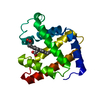 3h57C 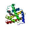 2mbwS C: citing same article ( S: Starting model for refinement |
|---|---|
| Similar structure data |


-Links
 PDBj
PDBj


- Assembly
| Deposited unit | 
| ||||||||
|---|---|---|---|---|---|---|---|---|---|
| 1 |
| ||||||||
| Unit cell |
|
-Components
| #1: Protein | Mass: 17355.146 Da / Num. of mol.: 1 / Mutation: H64L, V68N, D122N Source method: isolated from a genetically manipulated source Source: (gene. exp.)  |
|---|---|
| #2: Chemical | ChemComp-HEM /   Mass: 616.487 Da / Num. of mol.: 1 / Source method: obtained synthetically / Formula: C34H32FeN4O4 Mass: 616.487 Da / Num. of mol.: 1 / Source method: obtained synthetically / Formula: C34H32FeN4O4 |
| #3: Water | ChemComp-HOH /  Mass: 18.015 Da / Num. of mol.: 293 / Source method: isolated from a natural source / Formula: H2O Mass: 18.015 Da / Num. of mol.: 293 / Source method: isolated from a natural source / Formula: H2O |
-Experimental details
-Experiment
| Experiment | Method: X-RAY DIFFRACTION / Number of used crystals: 1 |
|---|
- Sample preparation
| Crystal | Density Matthews: 3.06 Å3/Da / Density % sol: 59.77 % |
|---|---|
| Crystal grow | Temperature: 293 K / Method: batch method / pH: 9 Details: Protein Solution (20mg/ml protein in 0.02M Tris.HCl pH9.0) mixed with mother liquor (3.2M Ammonium Sulphate, 0.05M Tris.HCl, pH9.0) for final concentrations of 2.5M Ammonium Sulphate, Batch ...Details: Protein Solution (20mg/ml protein in 0.02M Tris.HCl pH9.0) mixed with mother liquor (3.2M Ammonium Sulphate, 0.05M Tris.HCl, pH9.0) for final concentrations of 2.5M Ammonium Sulphate, Batch Method, temperature 293K |
-Data collection
| Diffraction | Mean temperature: 100 K |
|---|---|
| Diffraction source | Source: ROTATING ANODE / Type: RIGAKU RUH3R / Wavelength: 1.5418 Å |
| Detector | Type: RIGAKU RAXIS IV++ / Detector: IMAGE PLATE / Date: Apr 12, 2008 |
| Radiation | Monochromator: Multilayer / Protocol: SINGLE WAVELENGTH / Scattering type: x-ray |
| Radiation wavelength | Wavelength: 1.5418 Å / Relative weight: 1 |
| Reflection | Resolution: 1.54→39.03 Å / Num. all: 31355 / Num. obs: 26335 / % possible obs: 84 % / Observed criterion σ(I): 3 / Redundancy: 2.62 % / Biso Wilson estimate: 13 Å2 / Rmerge(I) obs: 0.035 / Χ2: 0.85 / Net I/σ(I): 18.9 / Scaling rejects: 522 |
| Reflection shell | Resolution: 1.54→1.59 Å / Redundancy: 1.04 % / Rmerge(I) obs: 0.111 / Mean I/σ(I) obs: 3 / Num. measured all: 580 / Num. unique all: 546 / Χ2: 2.8 / % possible all: 17.4 |
- Processing
| Software |
| ||||||||||||||||||||||||||||||||
|---|---|---|---|---|---|---|---|---|---|---|---|---|---|---|---|---|---|---|---|---|---|---|---|---|---|---|---|---|---|---|---|---|---|
| Refinement | Method to determine structure: FOURIER SYNTHESIS Starting model: 2mbw Resolution: 1.8→39.03 Å / Occupancy max: 1 / Occupancy min: 0.5 / FOM work R set: 0.871 / Isotropic thermal model: restrained / Cross valid method: THROUGHOUT / σ(F): 0 / Stereochemistry target values: Engh & Huber
| ||||||||||||||||||||||||||||||||
| Solvent computation | Bsol: 60.01 Å2 | ||||||||||||||||||||||||||||||||
| Displacement parameters | Biso max: 98.42 Å2 / Biso mean: 17.789 Å2 / Biso min: 4.17 Å2
| ||||||||||||||||||||||||||||||||
| Refine analyze |
| ||||||||||||||||||||||||||||||||
| Refinement step | Cycle: LAST / Resolution: 1.8→39.03 Å
| ||||||||||||||||||||||||||||||||
| Refine LS restraints |
| ||||||||||||||||||||||||||||||||
| LS refinement shell | Resolution: 1.8→1.91 Å / Rfactor Rfree error: 0.017
| ||||||||||||||||||||||||||||||||
| Xplor file |
|
