 Movie
Movie Controller

[English] 日本語
 Yorodumi
- PDB-3h2j: Crystal structure of the rice cell wall degrading esterase LipA f... -
+ Open data

- Basic information
| Entry | Database: PDB / ID: 3h2j | ||||||
|---|---|---|---|---|---|---|---|
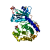
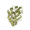
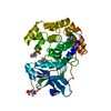
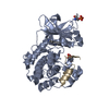
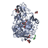
| Title | Crystal structure of the rice cell wall degrading esterase LipA from Xanthomonas oryzae | ||||||
 Components Components | esterase | ||||||
 Keywords Keywords | HYDROLASE / Xanthomonas oryzae pv. oryzae / esterase / cell wall degrading enzyme / rice / virulence / innate immune responses / pathogenesis / glycoside binding | ||||||
| Function / homology |  Function and homology information Function and homology informationtriacylglycerol lipase activity / aminopeptidase activity / lipid catabolic process Similarity search - Function | ||||||
| Biological species |  Xanthomonas oryzae pv. oryzae (bacteria) Xanthomonas oryzae pv. oryzae (bacteria) | ||||||
| Method |  X-RAY DIFFRACTION / MOLECULAR REPLACEMENT / Resolution: 1.89 Å X-RAY DIFFRACTION / MOLECULAR REPLACEMENT / Resolution: 1.89 Å | ||||||
 Authors Authors | Aparna, G. / Chatterjee, A. / Sonti, R.V. / Sankaranarayanan, R. | ||||||
 Citation Citation | Journal: Plant Cell / Year: 2009 Title: A Cell Wall-Degrading Esterase of Xanthomonas oryzae Requires a Unique Substrate Recognition Module for Pathogenesis on Rice Authors: Aparna, G. / Chatterjee, A. / Sonti, R.V. / Sankaranarayanan, R. | ||||||
| History |
|
- Structure visualization
| Structure viewer | Molecule: MolmilJmol/JSmol |
|---|
- Downloads & links
-Download
| PDBx/mmCIF format | 3h2j.cif.gz | 91.4 KB | Display | PDBx/mmCIF format |
|---|---|---|---|---|
| PDB format | pdb3h2j.ent.gz | 68.7 KB | Display | PDB format |
| PDBx/mmJSON format | 3h2j.json.gz | Tree view | PDBx/mmJSON format | |
| Others |  Other downloads Other downloads |
-Validation report
| Arichive directory | https://data.pdbj.org/pub/pdb/validation_reports/h2/3h2jftp://data.pdbj.org/pub/pdb/validation_reports/h2/3h2j | HTTPS FTP |
|---|
-Related structure data
| Related structure data | 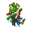 3h2gSC 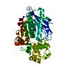 3h2hC 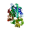 3h2iC 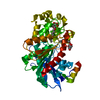 3h2kC S: Starting model for refinement C: citing same article ( |
|---|---|
| Similar structure data |


-Links
 PDBj
PDBj- Assembly
| Deposited unit | 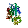
| ||||||||
|---|---|---|---|---|---|---|---|---|---|
| 1 |
| ||||||||
| Unit cell |
|
-Components
| #1: Protein | Mass: 42662.637 Da / Num. of mol.: 1 / Fragment: residues in UNP 45-441 Source method: isolated from a genetically manipulated source Source: (gene. exp.) Xanthomonas oryzae pv. oryzae (bacteria)Strain: BXO43 / Gene: LipA / Plasmid: pHM1 / Production host: Xanthomonas oryzae pv. oryzae (bacteria) / Strain (production host): BXO43References: UniProt: Q5H5J0, Hydrolases; Acting on ester bonds; Carboxylic-ester hydrolases |
|---|---|
| #2: Water | ChemComp-HOH /  Mass: 18.015 Da / Num. of mol.: 324 / Source method: isolated from a natural source / Formula: H2O Mass: 18.015 Da / Num. of mol.: 324 / Source method: isolated from a natural source / Formula: H2O |
| Has protein modification | Y |
-Experimental details
-Experiment
| Experiment | Method: X-RAY DIFFRACTION / Number of used crystals: 1 |
|---|
- Sample preparation
| Crystal | Density Matthews: 2.25 Å3/Da / Density % sol: 45.36 % |
|---|---|
| Crystal grow | Temperature: 298 K / Method: vapor diffusion, hanging drop / pH: 6 Details: 48% PEG 400, 0.10M MES, pH 6.0, VAPOR DIFFUSION, HANGING DROP, temperature 298K |
-Data collection
| Diffraction source | Source: ROTATING ANODE / Type: RIGAKU RUH3R / Wavelength: 1.5418 Å |
|---|---|
| Detector | Type: MAR scanner 345 mm plate / Detector: IMAGE PLATE / Date: Dec 18, 2006 |
| Radiation | Protocol: SINGLE WAVELENGTH / Monochromatic (M) / Laue (L): M / Scattering type: x-ray |
| Radiation wavelength | Wavelength: 1.5418 Å / Relative weight: 1 |
| Reflection | Resolution: 1.89→25 Å / Num. obs: 27294 / Biso Wilson estimate: 19.9 Å2 |
- Processing
| Software |
| ||||||||||||||||||||||||||||||||||||
|---|---|---|---|---|---|---|---|---|---|---|---|---|---|---|---|---|---|---|---|---|---|---|---|---|---|---|---|---|---|---|---|---|---|---|---|---|---|
| Refinement | Method to determine structure: MOLECULAR REPLACEMENT Starting model: 3H2G Resolution: 1.89→24.2 Å / Rfactor Rfree error: 0.006 / Data cutoff high absF: 1612603.41 / Data cutoff low absF: 0 / Isotropic thermal model: RESTRAINED / Cross valid method: THROUGHOUT / σ(F): 0
| ||||||||||||||||||||||||||||||||||||
| Solvent computation | Solvent model: FLAT MODEL / Bsol: 59.0006 Å2 / ksol: 0.361392 e/Å3 | ||||||||||||||||||||||||||||||||||||
| Displacement parameters | Biso mean: 22.8 Å2
| ||||||||||||||||||||||||||||||||||||
| Refine analyze |
| ||||||||||||||||||||||||||||||||||||
| Refinement step | Cycle: LAST / Resolution: 1.89→24.2 Å
| ||||||||||||||||||||||||||||||||||||
| Refine LS restraints |
| ||||||||||||||||||||||||||||||||||||
| LS refinement shell | Resolution: 1.89→1.96 Å / Rfactor Rfree error: 0.027 / Total num. of bins used: 10
| ||||||||||||||||||||||||||||||||||||
| Xplor file |
|
