[English] 日本語
 Yorodumi
- PDB-3h00: Structure of the C-terminal Domain of a Putative HIV-1 gp41 Fusio... -
+ Open data
- Basic information
| Entry | Database: PDB / ID: 3h00 | ||||||
|---|---|---|---|---|---|---|---|
| Title | Structure of the C-terminal Domain of a Putative HIV-1 gp41 Fusion Intermediate | ||||||
 Components Components | Envelope glycoprotein gp160 | ||||||
 Keywords Keywords | VIRAL PROTEIN / viral membrane fusion / HIV-1 / gp41 / envelope protein / neutralizing antibodies / AIDS / Apoptosis / Cell membrane / Cleavage on pair of basic residues / Disulfide bond / Fusion protein / Glycoprotein / Host-virus interaction / Lipoprotein / Membrane / Palmitate / Transmembrane / Viral immunoevasion / Virion | ||||||
| Function / homology |  Function and homology information Function and homology informationDectin-2 family / positive regulation of plasma membrane raft polarization / positive regulation of receptor clustering / host cell endosome membrane / clathrin-dependent endocytosis of virus by host cell / viral protein processing / fusion of virus membrane with host plasma membrane / fusion of virus membrane with host endosome membrane / viral envelope / virion attachment to host cell ...Dectin-2 family / positive regulation of plasma membrane raft polarization / positive regulation of receptor clustering / host cell endosome membrane / clathrin-dependent endocytosis of virus by host cell / viral protein processing / fusion of virus membrane with host plasma membrane / fusion of virus membrane with host endosome membrane / viral envelope / virion attachment to host cell / host cell plasma membrane / virion membrane / structural molecule activity / membrane Similarity search - Function | ||||||
| Biological species |  Human immunodeficiency virus type 1 lw12.3 isolate Human immunodeficiency virus type 1 lw12.3 isolate | ||||||
| Method |  X-RAY DIFFRACTION / X-RAY DIFFRACTION /  SYNCHROTRON / SYNCHROTRON /  MOLECULAR REPLACEMENT / Resolution: 2.2 Å MOLECULAR REPLACEMENT / Resolution: 2.2 Å | ||||||
 Authors Authors | Liu, J. | ||||||
 Citation Citation |  Journal: J.Virol. / Year: 2010 Journal: J.Virol. / Year: 2010Title: Role of a putative gp41 dimerization domain in human immunodeficiency virus type 1 membrane fusion. Authors: Liu, J. / Deng, Y. / Li, Q. / Dey, A.K. / Moore, J.P. / Lu, M. | ||||||
| History |
|
- Structure visualization
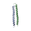
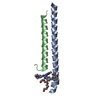
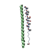
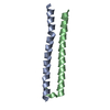
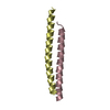
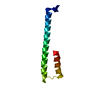
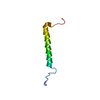
| Structure viewer | Molecule:  Molmil Molmil Jmol/JSmol Jmol/JSmol |
|---|
- Downloads & links
- Download
| PDBx/mmCIF format |  3h00.cif.gz 3h00.cif.gz | 67.7 KB | Display |  PDBx/mmCIF format PDBx/mmCIF format |
|---|---|---|---|---|
| PDB format |  pdb3h00.ent.gz pdb3h00.ent.gz | 51.8 KB | Display |  PDB format PDB format |
| PDBx/mmJSON format |  3h00.json.gz 3h00.json.gz | Tree view |  PDBx/mmJSON format PDBx/mmJSON format | |
| Others |  Other downloads Other downloads |
-Validation report
| Summary document |  3h00_validation.pdf.gz 3h00_validation.pdf.gz | 442.2 KB | Display |  wwPDB validaton report wwPDB validaton report |
|---|---|---|---|---|
| Full document |  3h00_full_validation.pdf.gz 3h00_full_validation.pdf.gz | 447.5 KB | Display | |
| Data in XML |  3h00_validation.xml.gz 3h00_validation.xml.gz | 9.3 KB | Display | |
| Data in CIF |  3h00_validation.cif.gz 3h00_validation.cif.gz | 12.3 KB | Display | |
| Arichive directory |  https://data.pdbj.org/pub/pdb/validation_reports/h0/3h00 https://data.pdbj.org/pub/pdb/validation_reports/h0/3h00 ftp://data.pdbj.org/pub/pdb/validation_reports/h0/3h00 ftp://data.pdbj.org/pub/pdb/validation_reports/h0/3h00 | HTTPS FTP |
-Related structure data
| Related structure data | 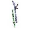 3gwoSC 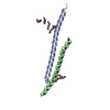 3h01C S: Starting model for refinement C: citing same article ( |
|---|---|
| Similar structure data |
- Links
Links
- Assembly
| Deposited unit | 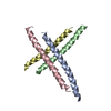
| ||||||||
|---|---|---|---|---|---|---|---|---|---|
| 1 | 
| ||||||||
| 2 | 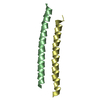
| ||||||||
| Unit cell |
|
- Components
| #1: Protein/peptide | Mass: 4796.156 Da / Num. of mol.: 4 / Fragment: C-TERMINAL DOMAIN (UNP residues 636 to 674) Source method: isolated from a genetically manipulated source Source: (gene. exp.)  Human immunodeficiency virus type 1 lw12.3 isolate Human immunodeficiency virus type 1 lw12.3 isolateStrain: HXB2 ISOLATE / Gene: env / Production host:  #2: Water | ChemComp-HOH / | |
|---|
-Experimental details
-Experiment
| Experiment | Method:  X-RAY DIFFRACTION / Number of used crystals: 1 X-RAY DIFFRACTION / Number of used crystals: 1 |
|---|
- Sample preparation
| Crystal | Density Matthews: 4.39 Å3/Da / Density % sol: 71.97 % |
|---|---|
| Crystal grow | Temperature: 295 K / Method: vapor diffusion, hanging drop / pH: 7.8 Details: 3% PEG8000, 0.1M Tris-HCl, 0.1M cesium chloride, pH 7.8, VAPOR DIFFUSION, HANGING DROP, temperature 295K |
-Data collection
| Diffraction | Mean temperature: 100 K | |||||||||||||||
|---|---|---|---|---|---|---|---|---|---|---|---|---|---|---|---|---|
| Diffraction source | Source:  SYNCHROTRON / Site: SYNCHROTRON / Site:  NSLS NSLS  / Beamline: X25 / Wavelength: 1.1 Å / Beamline: X25 / Wavelength: 1.1 Å | |||||||||||||||
| Detector | Type: ADSC QUANTUM 315 / Detector: CCD / Date: Aug 24, 2005 | |||||||||||||||
| Radiation | Protocol: SINGLE WAVELENGTH / Monochromatic (M) / Laue (L): M / Scattering type: x-ray | |||||||||||||||
| Radiation wavelength | Wavelength: 1.1 Å / Relative weight: 1 | |||||||||||||||
| Reflection twin |
| |||||||||||||||
| Reflection | Resolution: 2.2→40.1 Å / Num. all: 16151 / Num. obs: 16151 / % possible obs: 93.7 % / Observed criterion σ(F): 0 / Observed criterion σ(I): 0 / Redundancy: 3 % / Biso Wilson estimate: 46.5 Å2 / Rmerge(I) obs: 0.044 / Net I/σ(I): 18.7 | |||||||||||||||
| Reflection shell | Resolution: 2.2→2.28 Å / Redundancy: 2.5 % / Rmerge(I) obs: 0.269 / Mean I/σ(I) obs: 3.1 / Num. unique all: 1309 / % possible all: 76.2 |
- Processing
| Software |
| ||||||||||||||||||||||||||||||||||||||||||||||||||||||||||||||||||||||||||||||||||||||||||||||||||||||||||||||||||||||||||||||||||||||||||||||||||||||||||||||||||||||||||
|---|---|---|---|---|---|---|---|---|---|---|---|---|---|---|---|---|---|---|---|---|---|---|---|---|---|---|---|---|---|---|---|---|---|---|---|---|---|---|---|---|---|---|---|---|---|---|---|---|---|---|---|---|---|---|---|---|---|---|---|---|---|---|---|---|---|---|---|---|---|---|---|---|---|---|---|---|---|---|---|---|---|---|---|---|---|---|---|---|---|---|---|---|---|---|---|---|---|---|---|---|---|---|---|---|---|---|---|---|---|---|---|---|---|---|---|---|---|---|---|---|---|---|---|---|---|---|---|---|---|---|---|---|---|---|---|---|---|---|---|---|---|---|---|---|---|---|---|---|---|---|---|---|---|---|---|---|---|---|---|---|---|---|---|---|---|---|---|---|---|---|---|
| Refinement | Method to determine structure:  MOLECULAR REPLACEMENT MOLECULAR REPLACEMENTStarting model: PDB ENTRY 3GWO Resolution: 2.2→40.1 Å / Cor.coef. Fo:Fc: 0.949 / Cor.coef. Fo:Fc free: 0.938 / SU B: 9.299 / SU ML: 0.104 Isotropic thermal model: Isotropic with TLS groups assigned for each protein chain Cross valid method: THROUGHOUT / σ(F): 0 / σ(I): 0 / ESU R: 0.039 / ESU R Free: 0.037 / Stereochemistry target values: MAXIMUM LIKELIHOOD
| ||||||||||||||||||||||||||||||||||||||||||||||||||||||||||||||||||||||||||||||||||||||||||||||||||||||||||||||||||||||||||||||||||||||||||||||||||||||||||||||||||||||||||
| Solvent computation | Ion probe radii: 0.8 Å / Shrinkage radii: 0.8 Å / VDW probe radii: 1.4 Å / Solvent model: BABINET MODEL WITH MASK | ||||||||||||||||||||||||||||||||||||||||||||||||||||||||||||||||||||||||||||||||||||||||||||||||||||||||||||||||||||||||||||||||||||||||||||||||||||||||||||||||||||||||||
| Displacement parameters | Biso mean: 37.058 Å2
| ||||||||||||||||||||||||||||||||||||||||||||||||||||||||||||||||||||||||||||||||||||||||||||||||||||||||||||||||||||||||||||||||||||||||||||||||||||||||||||||||||||||||||
| Refinement step | Cycle: LAST / Resolution: 2.2→40.1 Å
| ||||||||||||||||||||||||||||||||||||||||||||||||||||||||||||||||||||||||||||||||||||||||||||||||||||||||||||||||||||||||||||||||||||||||||||||||||||||||||||||||||||||||||
| Refine LS restraints |
| ||||||||||||||||||||||||||||||||||||||||||||||||||||||||||||||||||||||||||||||||||||||||||||||||||||||||||||||||||||||||||||||||||||||||||||||||||||||||||||||||||||||||||
| LS refinement shell | Resolution: 2.196→2.253 Å / Total num. of bins used: 20
| ||||||||||||||||||||||||||||||||||||||||||||||||||||||||||||||||||||||||||||||||||||||||||||||||||||||||||||||||||||||||||||||||||||||||||||||||||||||||||||||||||||||||||
| Refinement TLS params. | Method: refined / Refine-ID: X-RAY DIFFRACTION
| ||||||||||||||||||||||||||||||||||||||||||||||||||||||||||||||||||||||||||||||||||||||||||||||||||||||||||||||||||||||||||||||||||||||||||||||||||||||||||||||||||||||||||
| Refinement TLS group |
|
 Movie
Movie Controller



 PDBj
PDBj


