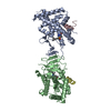
Entry Database : PDB / ID : 3gwxTitle MOLECULAR RECOGNITION OF FATTY ACIDS BY PEROXISOME PROLIFERATOR-ACTIVATED RECEPTORS PROTEIN (PEROXISOME PROLIFERATOR ACTIVATED RECEPTOR (PPAR-DELTA)) Keywords / / / / Function / homology Function Domain/homology Component
/ / / / / / / / / / / / / / / / / / / / / / / / / / / / / / / / / / / / / / / / / / / / / / / / / / / / / / / / / / / / / / / / / / / / / / / / / / / / / / / / / / / / / / / / / / / / / / / / / / / / / / / / / / / / / / / / / / / / / Biological species Homo sapiens (human)Method / / / Resolution : 2.4 Å Authors Xu, H.E. / Lambert, M.H. / Montana, V.G. / Parks, D.J. / Blanchard, S.G. / Brown, P.J. / Sternbach, D.D. / Lehmann, J.M. / Wisely, G.B. / Willson, T.M. ...Xu, H.E. / Lambert, M.H. / Montana, V.G. / Parks, D.J. / Blanchard, S.G. / Brown, P.J. / Sternbach, D.D. / Lehmann, J.M. / Wisely, G.B. / Willson, T.M. / Kliewer, S.A. / Milburn, M.V. Journal : Mol.Cell / Year : 1999Title : Molecular recognition of fatty acids by peroxisome proliferator-activated receptors.Authors : Xu, H.E. / Lambert, M.H. / Montana, V.G. / Parks, D.J. / Blanchard, S.G. / Brown, P.J. / Sternbach, D.D. / Lehmann, J.M. / Wisely, G.B. / Willson, T.M. / Kliewer, S.A. / Milburn, M.V. History Deposition Apr 26, 1999 Deposition site / Processing site Revision 1.0 Apr 26, 2000 Provider / Type Revision 1.1 Apr 26, 2008 Group Revision 1.2 Jul 13, 2011 Group Revision 1.3 Dec 27, 2023 Group / Database references / Derived calculationsCategory chem_comp_atom / chem_comp_bond ... chem_comp_atom / chem_comp_bond / database_2 / struct_site Item _database_2.pdbx_DOI / _database_2.pdbx_database_accession ... _database_2.pdbx_DOI / _database_2.pdbx_database_accession / _struct_site.pdbx_auth_asym_id / _struct_site.pdbx_auth_comp_id / _struct_site.pdbx_auth_seq_id
Show all Show less
 Movie
Movie Controller

 Yorodumi
 Open data
 Basic information
 Components
 Keywords
 Function and homology information
 Homo sapiens (human)
 X-RAY DIFFRACTION /
  Authors
 Citation
 Structure visualization
 Downloads & links
 Other downloads


 PDBj
PDBj
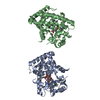
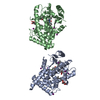
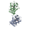
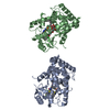
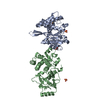
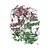
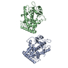
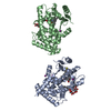
 Assembly


 Mass: 302.451 Da / Num. of mol.: 2 / Source method: obtained synthetically / Formula: C20H30O2
 Mass: 18.015 Da / Num. of mol.: 87 / Source method: isolated from a natural source / Formula: H2O
 Sample preparation
 / Beamline: 17-ID / Wavelength: 1.5418
  Processing