 Movie
Movie Controller

[English] 日本語
 Yorodumi
- PDB-3glc: Crystal Structure of E. coli LsrF in complex with Ribose-5-phosphate -
+ Open data

- Basic information
| Entry | Database: PDB / ID: 3glc | ||||||
|---|---|---|---|---|---|---|---|
| Title | Crystal Structure of E. coli LsrF in complex with Ribose-5-phosphate | ||||||
 Components Components | Aldolase lsrF | ||||||
 Keywords Keywords | LYASE / TIM barrel / Schiff base | ||||||
| Function / homology |  Function and homology information Function and homology information3-hydroxy-5-phosphooxypentane-2,4-dione thiolase / fructose-bisphosphate aldolase activity / acyltransferase activity, transferring groups other than amino-acyl groups / cytoplasm Similarity search - Function | ||||||
| Biological species |  | ||||||
| Method |  X-RAY DIFFRACTION / SYNCHROTRON / MOLECULAR REPLACEMENT / molecular replacement / Resolution: 2.5 Å X-RAY DIFFRACTION / SYNCHROTRON / MOLECULAR REPLACEMENT / molecular replacement / Resolution: 2.5 Å | ||||||
 Authors Authors | Miller, S.T. / Diaz, Z.C. | ||||||
 Citation Citation | Journal: Plos One / Year: 2009 Title: The crystal structure of the Escherichia coli autoinducer-2 processing protein LsrF. Authors: Diaz, Z. / Xavier, K.B. / Miller, S.T. | ||||||
| History |
|
- Structure visualization
| Structure viewer | Molecule: MolmilJmol/JSmol |
|---|
- Downloads & links
-Download
| PDBx/mmCIF format | 3glc.cif.gz | 1013.2 KB | Display | PDBx/mmCIF format |
|---|---|---|---|---|
| PDB format | pdb3glc.ent.gz | 850.3 KB | Display | PDB format |
| PDBx/mmJSON format | 3glc.json.gz | Tree view | PDBx/mmJSON format | |
| Others |  Other downloads Other downloads |
-Validation report
| Arichive directory | https://data.pdbj.org/pub/pdb/validation_reports/gl/3glcftp://data.pdbj.org/pub/pdb/validation_reports/gl/3glc | HTTPS FTP |
|---|
-Related structure data
| Related structure data |  3gkfSC 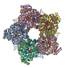 3gndC S: Starting model for refinement C: citing same article ( |
|---|---|
| Similar structure data |

-Links
 PDBj
PDBj
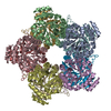
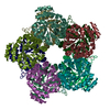
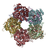
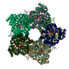
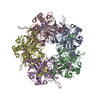
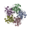
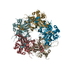
- Assembly
| Deposited unit | 
| ||||||||||||||||||||||||||||||||||||||||||||||||||||||||||||||||||||||||||||||||||||||||||||||||||||||||||||||||||||||||||||||
|---|---|---|---|---|---|---|---|---|---|---|---|---|---|---|---|---|---|---|---|---|---|---|---|---|---|---|---|---|---|---|---|---|---|---|---|---|---|---|---|---|---|---|---|---|---|---|---|---|---|---|---|---|---|---|---|---|---|---|---|---|---|---|---|---|---|---|---|---|---|---|---|---|---|---|---|---|---|---|---|---|---|---|---|---|---|---|---|---|---|---|---|---|---|---|---|---|---|---|---|---|---|---|---|---|---|---|---|---|---|---|---|---|---|---|---|---|---|---|---|---|---|---|---|---|---|---|---|
| 1 | 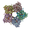
| ||||||||||||||||||||||||||||||||||||||||||||||||||||||||||||||||||||||||||||||||||||||||||||||||||||||||||||||||||||||||||||||
| 2 | 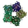
| ||||||||||||||||||||||||||||||||||||||||||||||||||||||||||||||||||||||||||||||||||||||||||||||||||||||||||||||||||||||||||||||
| Unit cell |
| ||||||||||||||||||||||||||||||||||||||||||||||||||||||||||||||||||||||||||||||||||||||||||||||||||||||||||||||||||||||||||||||
| Noncrystallographic symmetry (NCS) | NCS domain:
NCS domain segments: Component-ID: 1 / Ens-ID: 1 / Refine code: 1 / Auth seq-ID: -99999 - 99999 / Label seq-ID: -99999 - 99999
| ||||||||||||||||||||||||||||||||||||||||||||||||||||||||||||||||||||||||||||||||||||||||||||||||||||||||||||||||||||||||||||||
| Details | biological unit is a decamer. There are 2 biological units in the asymmetric unit (Chains ABCDEFGHIJ and chains KLMNOPQRST) |
-Components
| #1: Protein | Mass: 32322.145 Da / Num. of mol.: 20 / Fragment: Uncharacterized aldolase LsrF Source method: isolated from a genetically manipulated source Source: (gene. exp.) References: UniProt: P76143, Lyases; Carbon-carbon lyases; Aldehyde-lyases #2: Sugar | ChemComp-R5P /   Type: saccharide / Mass: 230.110 Da / Num. of mol.: 20 Type: saccharide / Mass: 230.110 Da / Num. of mol.: 20Source method: isolated from a genetically manipulated source Formula: C5H11O8P #3: Water | ChemComp-HOH / |  Mass: 18.015 Da / Num. of mol.: 334 / Source method: isolated from a natural source / Formula: H2O Mass: 18.015 Da / Num. of mol.: 334 / Source method: isolated from a natural source / Formula: H2O |
|---|
-Experimental details
-Experiment
| Experiment | Method: X-RAY DIFFRACTION / Number of used crystals: 1 |
|---|
- Sample preparation
| Crystal | Density Matthews: 2.3 Å3/Da / Density % sol: 46.52 % |
|---|---|
| Crystal grow | Temperature: 298 K / Method: vapor diffusion / pH: 7.5 Details: 4% PEG 400, 100 mM MgCl2, 2.3 M Ammonium Sulfate, pH 7.5, vapor diffusion, temperature 298K |
-Data collection
| Diffraction | Mean temperature: 100 K |
|---|---|
| Diffraction source | Source: SYNCHROTRON / Site: NSLS  / Beamline: X26C / Wavelength: 1 Å / Beamline: X26C / Wavelength: 1 Å |
| Detector | Type: ADSC QUANTUM 4 / Detector: CCD / Date: Oct 18, 2008 |
| Radiation | Monochromator: Si(111) / Protocol: SINGLE WAVELENGTH / Monochromatic (M) / Laue (L): M / Scattering type: x-ray |
| Radiation wavelength | Wavelength: 1 Å / Relative weight: 1 |
| Reflection | Resolution: 2.5→55.73 Å / Num. all: 327170 / Num. obs: 154484 / % possible obs: 81.7 % / Observed criterion σ(F): 0 / Observed criterion σ(I): 0 / Redundancy: 2.1 % / Biso Wilson estimate: 45.3 Å2 / Rmerge(I) obs: 0.065 / Net I/σ(I): 7.5 |
| Reflection shell | Resolution: 2.5→2.64 Å / Redundancy: 1.7 % / Rmerge(I) obs: 0.279 / Mean I/σ(I) obs: 1.8 / Num. unique all: 18764 / % possible all: 67.8 |
-Phasing
| Phasing | Method: molecular replacement |
|---|
- Processing
| Software |
| |||||||||||||||||||||||||||||||||||||||||||||||||||||||||||||||||||||||||||||||||||||||||||||||||||||||||||||||||
|---|---|---|---|---|---|---|---|---|---|---|---|---|---|---|---|---|---|---|---|---|---|---|---|---|---|---|---|---|---|---|---|---|---|---|---|---|---|---|---|---|---|---|---|---|---|---|---|---|---|---|---|---|---|---|---|---|---|---|---|---|---|---|---|---|---|---|---|---|---|---|---|---|---|---|---|---|---|---|---|---|---|---|---|---|---|---|---|---|---|---|---|---|---|---|---|---|---|---|---|---|---|---|---|---|---|---|---|---|---|---|---|---|---|---|
| Refinement | Method to determine structure: MOLECULAR REPLACEMENT Starting model: PDB entry 3GKF Resolution: 2.5→55.73 Å / Cor.coef. Fo:Fc: 0.934 / Cor.coef. Fo:Fc free: 0.911 / WRfactor Rfree: 0.235 / WRfactor Rwork: 0.208 / Occupancy max: 1 / Occupancy min: 1 / FOM work R set: 0.837 / SU B: 10.846 / SU ML: 0.238 / SU Rfree: 0.405 / Cross valid method: THROUGHOUT / σ(F): 0 / ESU R Free: 0.405 / Stereochemistry target values: MAXIMUM LIKELIHOOD / Details: U VALUES : REFINED INDIVIDUALLY
| |||||||||||||||||||||||||||||||||||||||||||||||||||||||||||||||||||||||||||||||||||||||||||||||||||||||||||||||||
| Solvent computation | Ion probe radii: 0.8 Å / Shrinkage radii: 0.8 Å / VDW probe radii: 1.4 Å / Solvent model: BABINET MODEL WITH MASK | |||||||||||||||||||||||||||||||||||||||||||||||||||||||||||||||||||||||||||||||||||||||||||||||||||||||||||||||||
| Displacement parameters | Biso max: 108.88 Å2 / Biso mean: 35.975 Å2 / Biso min: 5.43 Å2
| |||||||||||||||||||||||||||||||||||||||||||||||||||||||||||||||||||||||||||||||||||||||||||||||||||||||||||||||||
| Refinement step | Cycle: LAST / Resolution: 2.5→55.73 Å
| |||||||||||||||||||||||||||||||||||||||||||||||||||||||||||||||||||||||||||||||||||||||||||||||||||||||||||||||||
| Refine LS restraints |
| |||||||||||||||||||||||||||||||||||||||||||||||||||||||||||||||||||||||||||||||||||||||||||||||||||||||||||||||||
| Refine LS restraints NCS | Dom-ID: 1 / Ens-ID: 1 / Number: 2127 / Refine-ID: X-RAY DIFFRACTION
|