Mass: 18.015 Da / Num. of mol.: 458 / Source method: isolated from a natural source / Formula: H2O
Sequence details
THIS CONSTRUCT WAS EXPRESSED WITH A PURIFICATION TAG MGSDKIHHHHHHENLYFQG. THE TAG WAS REMOVED WITH ...THIS CONSTRUCT WAS EXPRESSED WITH A PURIFICATION TAG MGSDKIHHHHHHENLYFQG. THE TAG WAS REMOVED WITH TEV PROTEASE LEAVING ONLY A GLYCINE (0) FOLLOWED BY THE TARGET SEQUENCE.
-
Experimental details
-
Experiment
Experiment
Method: X-RAY DIFFRACTION / Number of used crystals: 1
-
Sample preparation
Crystal
Density Matthews: 2.18 Å3/Da / Density % sol: 43.47 %
Crystal grow
Temperature: 277 K / Method: vapor diffusion, sitting drop / pH: 8 Details: NANODROP, 59.1% 2-methyl-2,4-pentanediol, 0.1M Bicine pH 8.0, VAPOR DIFFUSION, SITTING DROP, temperature 277K
Resolution: 1.55→28.33 Å / Num. obs: 61448 / % possible obs: 92 % / Observed criterion σ(I): -3 / Biso Wilson estimate: 13.794 Å2 / Rmerge(I) obs: 0.057
Reflection shell
Resolution (Å)
Rmerge(I) obs
Mean I/σ(I) obs
Num. measured obs
Num. unique obs
Diffraction-ID
% possible all
1.55-1.61
0.52
1.5
14238
11275
1
85.2
1.61-1.67
0.418
1.9
13077
10381
1
90.7
1.67-1.75
0.337
2.4
14842
11791
1
91.2
1.75-1.84
0.233
3.3
13705
10954
1
91.9
1.84-1.95
0.165
4.6
13628
10919
1
92.5
1.95-2.1
0.102
6.7
14300
11479
1
92.9
2.1-2.31
0.067
9.7
14344
11566
1
93.3
2.31-2.65
0.052
12.2
14718
11856
1
94.2
2.65-3.33
0.032
18.3
14301
11558
1
94.2
3.33-28.33
0.018
28.8
14626
11810
1
94.4
-
Phasing
Phasing
Method: MAD
-
Processing
Software
Name
Version
Classification
NB
REFMAC
5.5.0053
refinement
PHENIX
refinement
SOLVE
phasing
MolProbity
3beta29
modelbuilding
XSCALE
datascaling
PDB_EXTRACT
3.006
dataextraction
MAR345
CCD
datacollection
XDS
datareduction
Refinement
Method to determine structure: MAD / Resolution: 1.55→28.33 Å / Cor.coef. Fo:Fc: 0.975 / Cor.coef. Fo:Fc free: 0.964 / Occupancy max: 1 / Occupancy min: 0.25 / SU B: 3.26 / SU ML: 0.05 / TLS residual ADP flag: LIKELY RESIDUAL / Cross valid method: THROUGHOUT / σ(F): 0 / ESU R: 0.068 / ESU R Free: 0.07 Stereochemistry target values: MAXIMUM LIKELIHOOD WITH PHASES Details: 1. HYDROGENS HAVE BEEN ADDED IN THE RIDING POSITIONS. 2. ATOM RECORDS CONTAIN RESIDUAL B FACTORS ONLY. 3. A MET-INHIBITION PROTOCOL WAS USED FOR SELENOMETHIONINE INCORPORATION DURING PROTEIN ...Details: 1. HYDROGENS HAVE BEEN ADDED IN THE RIDING POSITIONS. 2. ATOM RECORDS CONTAIN RESIDUAL B FACTORS ONLY. 3. A MET-INHIBITION PROTOCOL WAS USED FOR SELENOMETHIONINE INCORPORATION DURING PROTEIN EXPRESSION. THE OCCUPANCY OF THE SE ATOMS IN THE MSE RESIDUES WAS REDUCED TO 0.75 FOR THE REDUCED SCATTERING POWER DUE TO PARTIAL S-MET INCORPORATION. 4. 2-METHYL-2,4-PENTANEDIOL (MPD) FROM THE CRYSTALLIZATION CONDITION HAS BEEN MODELED IN THE SOLVENT STRUCTURE. 5. PYRIDOXAL PHOSPHATE IN DUAL CONFORMATION AS FREE PLP AND COVALENTLY ATTACHED TO LYS289 (LLP) HAS BEEN MODELED AT THE PUTATIVE ACTIVE SITE BASED ON DIFFERENCE DENSITY AND OMIT MAPS AND STRUCTURAL SIMILARITY TO PLP-DEPENDENT AMINOTRANSFERASES. 6. RESIDUE ALA288 IS PRESENT IN THE VICINITY OF THE PUTATIVE ACTIVE SITE AND IS A RAMACHANDRAN OUTLIER. THIS COULD HAVE SOME FUNCTIONAL SIGNIFICANCE.
Rfactor
Num. reflection
% reflection
Selection details
Rfree
0.168
3122
5.1 %
RANDOM
Rwork
0.139
-
-
-
obs
0.14
61444
98.32 %
-
Solvent computation
Ion probe radii: 0.8 Å / Shrinkage radii: 0.8 Å / VDW probe radii: 1.2 Å / Solvent model: MASK
In the structure databanks used in Yorodumi, some data are registered as the other names, "COVID-19 virus" and "2019-nCoV". Here are the details of the virus and the list of structure data.
Jan 31, 2019. EMDB accession codes are about to change! (news from PDBe EMDB page)
EMDB accession codes are about to change! (news from PDBe EMDB page)
The allocation of 4 digits for EMDB accession codes will soon come to an end. Whilst these codes will remain in use, new EMDB accession codes will include an additional digit and will expand incrementally as the available range of codes is exhausted. The current 4-digit format prefixed with “EMD-” (i.e. EMD-XXXX) will advance to a 5-digit format (i.e. EMD-XXXXX), and so on. It is currently estimated that the 4-digit codes will be depleted around Spring 2019, at which point the 5-digit format will come into force.
The EM Navigator/Yorodumi systems omit the EMD- prefix.
Related info.:Q: What is EMD? / ID/Accession-code notation in Yorodumi/EM Navigator
Yorodumi is a browser for structure data from EMDB, PDB, SASBDB, etc.
This page is also the successor to EM Navigator detail page, and also detail information page/front-end page for Omokage search.
The word "yorodu" (or yorozu) is an old Japanese word meaning "ten thousand". "mi" (miru) is to see.
Related info.:EMDB / PDB / SASBDB / Comparison of 3 databanks / Yorodumi Search / Aug 31, 2016. New EM Navigator & Yorodumi / Yorodumi Papers / Jmol/JSmol / Function and homology information / Changes in new EM Navigator and Yorodumi
 Movie
Movie Controller

 Yorodumi
 Open data
 Basic information
 Components
 Keywords
 Function and homology information
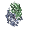
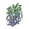
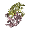
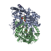
 Mesorhizobium loti (bacteria)
 X-RAY DIFFRACTION /
  Authors
 Citation
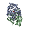
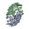
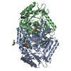
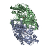
 Structure visualization
 Downloads & links
 Other downloads
 PDBj
PDBj Assembly


 Mass: 247.142 Da / Num. of mol.: 1 / Source method: obtained synthetically / Formula: C8H10NO6P
 Mass: 118.174 Da / Num. of mol.: 2 / Source method: obtained synthetically / Formula: C6H14O2 / Comment: precipitant*YM
 Mass: 18.015 Da / Num. of mol.: 458 / Source method: isolated from a natural source / Formula: H2O
 Sample preparation
 / Beamline: BL9-2 / Wavelength: 0.91162, 0.97941, 0.97954
 Processing