 Movie
Movie Controller

[English] 日本語
 Yorodumi
- PDB-3gfh: Crystal structure of EUTL shell protein of the bacterial ethanola... -
+ Open data

- Basic information
| Entry | Database: PDB / ID: 3gfh | ||||||
|---|---|---|---|---|---|---|---|
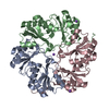
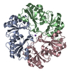
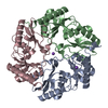
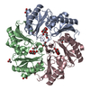
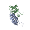
| Title | Crystal structure of EUTL shell protein of the bacterial ethanolamine micrompartment | ||||||
 Components Components | Ethanolamine utilization protein eutL | ||||||
 Keywords Keywords | STRUCTURAL PROTEIN / bacterial mircocompartment / shell protein / ethanolamine | ||||||
| Function / homology |  Function and homology information Function and homology informationethanolamine degradation polyhedral organelle / ethanolamine catabolic process / structural molecule activity / zinc ion binding / identical protein binding Similarity search - Function | ||||||
| Biological species |  | ||||||
| Method |  X-RAY DIFFRACTION / SYNCHROTRON / SAD / Resolution: 2.2 Å X-RAY DIFFRACTION / SYNCHROTRON / SAD / Resolution: 2.2 Å | ||||||
 Authors Authors | Sagermann, M. / Nikolakakis, K. / Ohtaki, A. | ||||||
 Citation Citation | Journal: Proc.Natl.Acad.Sci.USA / Year: 2009 Title: Crystal structure of the EutL shell protein of the ethanolamine ammonia lyase microcompartment Authors: Sagermann, M. / Ohtaki, A. / Nikolakakis, K. | ||||||
| History |
|
- Structure visualization
| Structure viewer | Molecule: MolmilJmol/JSmol |
|---|
- Downloads & links
-Download
| PDBx/mmCIF format | 3gfh.cif.gz | 92.5 KB | Display | PDBx/mmCIF format |
|---|---|---|---|---|
| PDB format | pdb3gfh.ent.gz | 71.6 KB | Display | PDB format |
| PDBx/mmJSON format | 3gfh.json.gz | Tree view | PDBx/mmJSON format | |
| Others |  Other downloads Other downloads |
-Validation report
| Summary document | 3gfh_validation.pdf.gz | 449.5 KB | Display | wwPDB validaton report |
|---|---|---|---|---|
| Full document | 3gfh_full_validation.pdf.gz | 481.2 KB | Display | |
| Data in XML | 3gfh_validation.xml.gz | 23.8 KB | Display | |
| Data in CIF | 3gfh_validation.cif.gz | 32.9 KB | Display | |
| Arichive directory | https://data.pdbj.org/pub/pdb/validation_reports/gf/3gfhftp://data.pdbj.org/pub/pdb/validation_reports/gf/3gfh | HTTPS FTP |
-Related structure data
| Related structure data | |
|---|---|
| Similar structure data |


-Links
 PDBj
PDBj


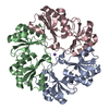
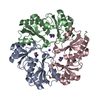
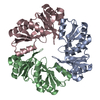
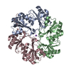
- Assembly
| Deposited unit | 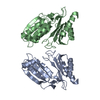
| ||||||||
|---|---|---|---|---|---|---|---|---|---|
| 1 | 
| ||||||||
| 2 | 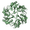
| ||||||||
| Unit cell |
|
-Components
| #1: Protein | Mass: 23633.656 Da / Num. of mol.: 2 Source method: isolated from a genetically manipulated source Source: (gene. exp.) #2: Chemical |   Mass: 200.590 Da / Num. of mol.: 2 / Source method: obtained synthetically / Formula: Hg Mass: 200.590 Da / Num. of mol.: 2 / Source method: obtained synthetically / Formula: Hg#3: Water | ChemComp-HOH / |  Mass: 18.015 Da / Num. of mol.: 171 / Source method: isolated from a natural source / Formula: H2O Mass: 18.015 Da / Num. of mol.: 171 / Source method: isolated from a natural source / Formula: H2O |
|---|
-Experimental details
-Experiment
| Experiment | Method: X-RAY DIFFRACTION / Number of used crystals: 1 |
|---|
- Sample preparation
| Crystal | Density Matthews: 2.29 Å3/Da / Density % sol: 46.27 % |
|---|---|
| Crystal grow | Temperature: 298 K / pH: 6.5 Details: 2M Nacl, 100mM phosphate, MES buffer pH6.5, 5% PEG 400, VAPOR DIFFUSION, HANGING DROP, temperature 298K |
-Data collection
| Diffraction | Mean temperature: 100 K | |||||||||||||||
|---|---|---|---|---|---|---|---|---|---|---|---|---|---|---|---|---|
| Diffraction source | Source: SYNCHROTRON / Site: SSRL  / Beamline: BL9-1 / Wavelength: 0.97946 / Beamline: BL9-1 / Wavelength: 0.97946 | |||||||||||||||
| Detector | Type: ADSC QUANTUM 315r / Detector: CCD / Details: SI-MIRRORS | |||||||||||||||
| Radiation | Protocol: SINGLE WAVELENGTH / Monochromatic (M) / Laue (L): M / Scattering type: x-ray | |||||||||||||||
| Radiation wavelength | Wavelength: 0.97946 Å / Relative weight: 1 | |||||||||||||||
| Reflection twin |
| |||||||||||||||
| Reflection | Resolution: 1.95→19.95 Å / Num. obs: 51500 / % possible obs: 85.8 % / Observed criterion σ(I): 0 / Biso Wilson estimate: 27.3 Å2 / Rmerge(I) obs: 0.08 | |||||||||||||||
| Reflection shell | Resolution: 1.95→2.3 Å / Rmerge(I) obs: 0.278 / Mean I/σ(I) obs: 4.46 / % possible all: 74.3 |
- Processing
| Software |
| ||||||||||||||||||||||||||||||||||||||||||||||||||||||||||||||||||||||||||||||||||||||||||||||||||||||||||||||||||||||||||||||||||||||||||||||||||||||||||||||||||||||||||
|---|---|---|---|---|---|---|---|---|---|---|---|---|---|---|---|---|---|---|---|---|---|---|---|---|---|---|---|---|---|---|---|---|---|---|---|---|---|---|---|---|---|---|---|---|---|---|---|---|---|---|---|---|---|---|---|---|---|---|---|---|---|---|---|---|---|---|---|---|---|---|---|---|---|---|---|---|---|---|---|---|---|---|---|---|---|---|---|---|---|---|---|---|---|---|---|---|---|---|---|---|---|---|---|---|---|---|---|---|---|---|---|---|---|---|---|---|---|---|---|---|---|---|---|---|---|---|---|---|---|---|---|---|---|---|---|---|---|---|---|---|---|---|---|---|---|---|---|---|---|---|---|---|---|---|---|---|---|---|---|---|---|---|---|---|---|---|---|---|---|---|---|
| Refinement | Method to determine structure: SAD Starting model: MODEL WAS DERIVED FROM FITTING INTO A 3.5 A SAD DENSITY DERIVED FROM TWO MERCURY ATOMS. Resolution: 2.2→19.65 Å / Cor.coef. Fo:Fc: 0.906 / Cor.coef. Fo:Fc free: 0.866 / SU B: 9.162 / SU ML: 0.228 / Cross valid method: THROUGHOUT / σ(F): 0 / ESU R: 0.034 / ESU R Free: 0.033 / Stereochemistry target values: MAXIMUM LIKELIHOOD Details: THE STRUCTURE WAS REFINED AGAINST TWINNED DATA AS PUBLISHED. The data is hemohedral twinning with twinning operators: (h,-h-k,-l) and corresponding twinned fractions: 0.575, 0.425. RESIDUES ...Details: THE STRUCTURE WAS REFINED AGAINST TWINNED DATA AS PUBLISHED. The data is hemohedral twinning with twinning operators: (h,-h-k,-l) and corresponding twinned fractions: 0.575, 0.425. RESIDUES 2-216 COULD BE FITTED RELIABLY INTO THE ELECTRON DENSITY MAP. HYDROGENS HAVE BEEN ADDED IN THE RIDING POSITIONS.
| ||||||||||||||||||||||||||||||||||||||||||||||||||||||||||||||||||||||||||||||||||||||||||||||||||||||||||||||||||||||||||||||||||||||||||||||||||||||||||||||||||||||||||
| Solvent computation | Ion probe radii: 0.8 Å / Shrinkage radii: 0.8 Å / VDW probe radii: 1.4 Å / Solvent model: BABINET MODEL WITH MASK | ||||||||||||||||||||||||||||||||||||||||||||||||||||||||||||||||||||||||||||||||||||||||||||||||||||||||||||||||||||||||||||||||||||||||||||||||||||||||||||||||||||||||||
| Displacement parameters | Biso mean: 19.34 Å2
| ||||||||||||||||||||||||||||||||||||||||||||||||||||||||||||||||||||||||||||||||||||||||||||||||||||||||||||||||||||||||||||||||||||||||||||||||||||||||||||||||||||||||||
| Refinement step | Cycle: LAST / Resolution: 2.2→19.65 Å
| ||||||||||||||||||||||||||||||||||||||||||||||||||||||||||||||||||||||||||||||||||||||||||||||||||||||||||||||||||||||||||||||||||||||||||||||||||||||||||||||||||||||||||
| Refine LS restraints |
| ||||||||||||||||||||||||||||||||||||||||||||||||||||||||||||||||||||||||||||||||||||||||||||||||||||||||||||||||||||||||||||||||||||||||||||||||||||||||||||||||||||||||||
| LS refinement shell | Resolution: 2.2→2.26 Å / Total num. of bins used: 20
|
