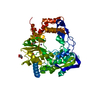
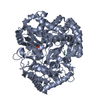
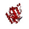
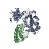
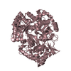
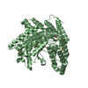
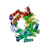
- PDB-3gdi: Mammalian Clock Protein mPER2 - Crystal Structure of a PAS Domain... -
+
データを開く
IDまたはキーワード:
読み込み中...
-
基本情報
登録情報
データベース: PDB / ID: 3gdi
タイトル
Mammalian Clock Protein mPER2 - Crystal Structure of a PAS Domain Fragment
要素
Period circadian protein homolog 2
キーワード
TRANSCRIPTION / tandem PAS domains / Biological rhythms / Nucleus / Phosphoprotein / Transcription regulation
機能・相同性
機能・相同性情報
regulation of glutamate uptake involved in transmission of nerve impulse / Cry-Per complex / negative regulation of termination of DNA-templated transcription / negative regulation of fat cell proliferation / negative regulation of circadian rhythm / lactate biosynthetic process / histone methyltransferase binding / glycogen biosynthetic process / pre-mRNA binding / RNA polymerase binding ...regulation of glutamate uptake involved in transmission of nerve impulse / Cry-Per complex / negative regulation of termination of DNA-templated transcription / negative regulation of fat cell proliferation / negative regulation of circadian rhythm / lactate biosynthetic process / histone methyltransferase binding / glycogen biosynthetic process / pre-mRNA binding / RNA polymerase binding / white fat cell differentiation / regulation of neurogenesis / regulation of vasoconstriction / transcription regulator inhibitor activity / negative regulation of protein ubiquitination / regulation of insulin secretion / response to ischemia / nuclear receptor binding / transcription corepressor binding / gluconeogenesis / circadian regulation of gene expression / fatty acid metabolic process / regulation of circadian rhythm / circadian rhythm / kinase binding / histone deacetylase binding / positive regulation of cold-induced thermogenesis / DNA-binding transcription factor binding / transcription coactivator activity / transcription cis-regulatory region binding / regulation of cell cycle / chromatin remodeling / RNA polymerase II cis-regulatory region sequence-specific DNA binding / negative regulation of DNA-templated transcription / perinuclear region of cytoplasm / negative regulation of transcription by RNA polymerase II / nucleoplasm / identical protein binding / nucleus / cytosol / cytoplasm 類似検索 - 分子機能
Period circadian protein homolog bHLH-like domain / Period circadian-like, C-terminal / : / Period circadian-like, C-terminal / Period circadian protein homolog 3-like, PAS-A domain / : / PAS fold-3 / PAS fold / PAS domain / Beta-Lactamase ...Period circadian protein homolog bHLH-like domain / Period circadian-like, C-terminal / : / Period circadian-like, C-terminal / Period circadian protein homolog 3-like, PAS-A domain / : / PAS fold-3 / PAS fold / PAS domain / Beta-Lactamase / PAS domain / PAS repeat profile. / PAS domain / PAS domain superfamily / 2-Layer Sandwich / Alpha Beta 類似検索 - ドメイン・相同性
 ムービー
ムービー コントローラー


 データを開く
 基本情報
基本情報 要素
要素 キーワード
キーワード 機能・相同性情報
 X線回折 /
  データ登録者
 引用
引用 構造の表示
構造の表示 ダウンロードとリンク
 その他のダウンロード
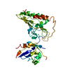


 PDBj
PDBj

 集合体
集合体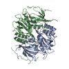

 分子量: 18.015 Da / 分子数: 198 / 由来タイプ: 天然 / 式: H2O
 試料調製
試料調製 / ビームライン: X06SA / 波長: 0.97805 Å
 解析
解析