- PDB-3gdi: Mammalian Clock Protein mPER2 - Crystal Structure of a PAS Domain... -
+
Open data
ID or keywords:
Loading...
-
Basic information
Entry
Database: PDB / ID: 3gdi
Title
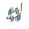
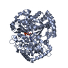
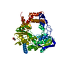
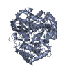
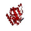
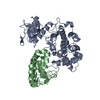
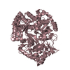
Mammalian Clock Protein mPER2 - Crystal Structure of a PAS Domain Fragment
Components
Period circadian protein homolog 2
Keywords
TRANSCRIPTION / tandem PAS domains / Biological rhythms / Nucleus / Phosphoprotein / Transcription regulation
Function / homology
Function and homology information
regulation of glutamate uptake involved in transmission of nerve impulse / Cry-Per complex / negative regulation of termination of DNA-templated transcription / negative regulation of fat cell proliferation / Degradation of CRY and PER proteins / negative regulation of circadian rhythm / lactate biosynthetic process / histone methyltransferase binding / glycogen biosynthetic process / pre-mRNA binding ...regulation of glutamate uptake involved in transmission of nerve impulse / Cry-Per complex / negative regulation of termination of DNA-templated transcription / negative regulation of fat cell proliferation / Degradation of CRY and PER proteins / negative regulation of circadian rhythm / lactate biosynthetic process / histone methyltransferase binding / glycogen biosynthetic process / pre-mRNA binding / RNA polymerase binding / entrainment of circadian clock by photoperiod / regulation of insulin secretion / white fat cell differentiation / regulation of neurogenesis / regulation of vasoconstriction / transcription regulator inhibitor activity / negative regulation of protein ubiquitination / response to ischemia / nuclear receptor binding / transcription corepressor binding / gluconeogenesis / circadian regulation of gene expression / circadian rhythm / fatty acid metabolic process / regulation of circadian rhythm / histone deacetylase binding / kinase binding / positive regulation of cold-induced thermogenesis / DNA-binding transcription factor binding / transcription coactivator activity / regulation of cell cycle / transcription cis-regulatory region binding / RNA polymerase II cis-regulatory region sequence-specific DNA binding / chromatin remodeling / negative regulation of DNA-templated transcription / perinuclear region of cytoplasm / negative regulation of transcription by RNA polymerase II / nucleoplasm / identical protein binding / nucleus / cytoplasm / cytosol Similarity search - Function
: / Period circadian protein homolog bHLH-like domain / Period circadian-like, C-terminal / : / Period circadian-like, C-terminal / Period circadian protein homolog 3-like, PAS-A domain / : / PAS fold-3 / PAS fold / PAS domain ...: / Period circadian protein homolog bHLH-like domain / Period circadian-like, C-terminal / : / Period circadian-like, C-terminal / Period circadian protein homolog 3-like, PAS-A domain / : / PAS fold-3 / PAS fold / PAS domain / Beta-Lactamase / PAS domain / PAS repeat profile. / PAS domain / PAS domain superfamily / 2-Layer Sandwich / Alpha Beta Similarity search - Domain/homology
In the structure databanks used in Yorodumi, some data are registered as the other names, "COVID-19 virus" and "2019-nCoV". Here are the details of the virus and the list of structure data.
Jan 31, 2019. EMDB accession codes are about to change! (news from PDBe EMDB page)
EMDB accession codes are about to change! (news from PDBe EMDB page)
The allocation of 4 digits for EMDB accession codes will soon come to an end. Whilst these codes will remain in use, new EMDB accession codes will include an additional digit and will expand incrementally as the available range of codes is exhausted. The current 4-digit format prefixed with “EMD-” (i.e. EMD-XXXX) will advance to a 5-digit format (i.e. EMD-XXXXX), and so on. It is currently estimated that the 4-digit codes will be depleted around Spring 2019, at which point the 5-digit format will come into force.
The EM Navigator/Yorodumi systems omit the EMD- prefix.
Related info.:Q: What is EMD? / ID/Accession-code notation in Yorodumi/EM Navigator
Yorodumi is a browser for structure data from EMDB, PDB, SASBDB, etc.
This page is also the successor to EM Navigator detail page, and also detail information page/front-end page for Omokage search.
The word "yorodu" (or yorozu) is an old Japanese word meaning "ten thousand". "mi" (miru) is to see.
Related info.:EMDB / PDB / SASBDB / Comparison of 3 databanks / Yorodumi Search / Aug 31, 2016. New EM Navigator & Yorodumi / Yorodumi Papers / Jmol/JSmol / Function and homology information / Changes in new EM Navigator and Yorodumi
 Movie
Movie Controller

 Yorodumi
 Open data
 Basic information
 Components
 Keywords
 Function and homology information
 X-RAY DIFFRACTION /
  Authors
 Citation
 Structure visualization
 Downloads & links
 Other downloads
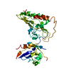


 PDBj
PDBj

 Assembly

 Mass: 18.015 Da / Num. of mol.: 198 / Source method: isolated from a natural source / Formula: H2O
 Sample preparation
 / Beamline: X06SA / Wavelength: 0.97805 Å
 Processing