 Movie
Movie Controller

[English] 日本語
 Yorodumi
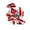
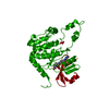
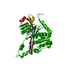
- PDB-3g7v: Islet Amyloid Polypeptide (IAPP or Amylin) fused to Maltose Bindi... -
+ Open data

- Basic information
| Entry | Database: PDB / ID: 3g7v | |||||||||
|---|---|---|---|---|---|---|---|---|---|---|
| Title | Islet Amyloid Polypeptide (IAPP or Amylin) fused to Maltose Binding Protein | |||||||||
 Components Components | Maltose-binding periplasmic protein, Islet amyloid polypeptide fusion protein | |||||||||
 Keywords Keywords | Sugar binding protein / Hormone / native fold for amyloidogenic protein / Sugar transport / Transport / Amidation / Amyloid / Cleavage on pair of basic residues / Secreted | |||||||||
| Function / homology |  Function and homology information Function and homology informationamylin receptor 3 signaling pathway / amylin receptor 2 signaling pathway / amylin receptor 1 signaling pathway / amylin receptor signaling pathway / Calcitonin-like ligand receptors / negative regulation of amyloid fibril formation / negative regulation of bone resorption / eating behavior / negative regulation of osteoclast differentiation / detection of maltose stimulus ...amylin receptor 3 signaling pathway / amylin receptor 2 signaling pathway / amylin receptor 1 signaling pathway / amylin receptor signaling pathway / Calcitonin-like ligand receptors / negative regulation of amyloid fibril formation / negative regulation of bone resorption / eating behavior / negative regulation of osteoclast differentiation / detection of maltose stimulus / Regulation of gene expression in beta cells / maltose transport complex / carbohydrate transport / positive regulation of cAMP/PKA signal transduction / carbohydrate transmembrane transporter activity / bone resorption / maltose binding / maltose transport / maltodextrin transmembrane transport / negative regulation of protein-containing complex assembly / ATP-binding cassette (ABC) transporter complex, substrate-binding subunit-containing / positive regulation of calcium-mediated signaling / ATP-binding cassette (ABC) transporter complex / osteoclast differentiation / sensory perception of pain / cell chemotaxis / hormone activity / cell-cell signaling / amyloid-beta binding / outer membrane-bounded periplasmic space / G alpha (s) signalling events / periplasmic space / positive regulation of MAPK cascade / positive regulation of apoptotic process / Amyloid fiber formation / receptor ligand activity / signaling receptor binding / neuronal cell body / apoptotic process / DNA damage response / lipid binding / signal transduction / : / extracellular region / membrane / identical protein binding Similarity search - Function | |||||||||
| Biological species |   Homo sapiens (human) Homo sapiens (human) | |||||||||
| Method |  X-RAY DIFFRACTION / SYNCHROTRON / MOLECULAR REPLACEMENT / molecular replacement / Resolution: 1.86 Å X-RAY DIFFRACTION / SYNCHROTRON / MOLECULAR REPLACEMENT / molecular replacement / Resolution: 1.86 Å | |||||||||
 Authors Authors | Wiltzius, J.J.W. / Sawaya, M.R. / Eisenberg, D. | |||||||||
 Citation Citation | Journal: Protein Sci. / Year: 2009 Title: Atomic structures of IAPP (amylin) fusions suggest a mechanism for fibrillation and the role of insulin in the process Authors: Wiltzius, J.J. / Sievers, S.A. / Sawaya, M.R. / Eisenberg, D. | |||||||||
| History |
|
- Structure visualization
| Structure viewer | Molecule: MolmilJmol/JSmol |
|---|
- Downloads & links
-Download
| PDBx/mmCIF format | 3g7v.cif.gz | 330.3 KB | Display | PDBx/mmCIF format |
|---|---|---|---|---|
| PDB format | pdb3g7v.ent.gz | 268 KB | Display | PDB format |
| PDBx/mmJSON format | 3g7v.json.gz | Tree view | PDBx/mmJSON format | |
| Others |  Other downloads Other downloads |
-Validation report
| Arichive directory | https://data.pdbj.org/pub/pdb/validation_reports/g7/3g7vftp://data.pdbj.org/pub/pdb/validation_reports/g7/3g7v | HTTPS FTP |
|---|
-Related structure data
| Related structure data | 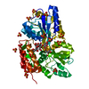 3g7wC 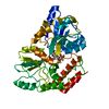 1mpdS C: citing same article ( S: Starting model for refinement |
|---|---|
| Similar structure data |


-Links
 PDBj
PDBj


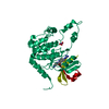
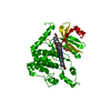
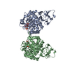
- Assembly
| Deposited unit | 
| ||||||||
|---|---|---|---|---|---|---|---|---|---|
| 1 | 
| ||||||||
| 2 | 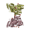
| ||||||||
| Unit cell |
|
-Components
| #1: Protein | Mass: 44442.172 Da / Num. of mol.: 4 Source method: isolated from a genetically manipulated source Source: (gene. exp.) Homo sapiens (human)Gene: malE, b4034, JW3994, IAPP / Plasmid: pMal-a1 / Production host: #2: Polysaccharide | alpha-D-glucopyranose-(1-4)-alpha-D-glucopyranose / alpha-maltose   Source method: isolated from a genetically manipulated source Details: oligosaccharide / References: alpha-maltose #3: Chemical | ChemComp-SO4 /   Mass: 96.063 Da / Num. of mol.: 15 / Source method: obtained synthetically / Formula: SO4 Mass: 96.063 Da / Num. of mol.: 15 / Source method: obtained synthetically / Formula: SO4#4: Chemical | ChemComp-GOL /   Mass: 92.094 Da / Num. of mol.: 5 / Source method: obtained synthetically / Formula: C3H8O3 Mass: 92.094 Da / Num. of mol.: 5 / Source method: obtained synthetically / Formula: C3H8O3#5: Water | ChemComp-HOH / |  Mass: 18.015 Da / Num. of mol.: 862 / Source method: isolated from a natural source / Formula: H2O Mass: 18.015 Da / Num. of mol.: 862 / Source method: isolated from a natural source / Formula: H2O |
|---|
-Experimental details
-Experiment
| Experiment | Method: X-RAY DIFFRACTION / Number of used crystals: 1 |
|---|
- Sample preparation
| Crystal | Density Matthews: 2.93 Å3/Da / Density % sol: 58.07 % |
|---|---|
| Crystal grow | Temperature: 298 K / Method: vapor diffusion, hanging drop / pH: 4.6 Details: 2.0 M Ammonium Sulfate, 0.1M Sodium Acetate pH 4.6, vapor diffusion, hanging drop, temperature 298K |
-Data collection
| Diffraction | Mean temperature: 100 K | |||||||||||||||||||||||||||||||||||||||||||||||||||||||||||||||||||||||||||||
|---|---|---|---|---|---|---|---|---|---|---|---|---|---|---|---|---|---|---|---|---|---|---|---|---|---|---|---|---|---|---|---|---|---|---|---|---|---|---|---|---|---|---|---|---|---|---|---|---|---|---|---|---|---|---|---|---|---|---|---|---|---|---|---|---|---|---|---|---|---|---|---|---|---|---|---|---|---|---|
| Diffraction source | Source: SYNCHROTRON / Site: ALS  / Beamline: 8.2.2 / Wavelength: 1 Å / Beamline: 8.2.2 / Wavelength: 1 Å | |||||||||||||||||||||||||||||||||||||||||||||||||||||||||||||||||||||||||||||
| Detector | Type: ADSC QUANTUM 315 / Detector: CCD / Date: Apr 28, 2007 | |||||||||||||||||||||||||||||||||||||||||||||||||||||||||||||||||||||||||||||
| Radiation | Protocol: SINGLE WAVELENGTH / Monochromatic (M) / Laue (L): M / Scattering type: x-ray | |||||||||||||||||||||||||||||||||||||||||||||||||||||||||||||||||||||||||||||
| Radiation wavelength | Wavelength: 1 Å / Relative weight: 1 | |||||||||||||||||||||||||||||||||||||||||||||||||||||||||||||||||||||||||||||
| Reflection | Resolution: 1.85→90 Å / Num. all: 169517 / Num. obs: 169517 / % possible obs: 98.4 % / Observed criterion σ(I): -3 / Redundancy: 5.5 % / Biso Wilson estimate: 35.6 Å2 / Rmerge(I) obs: 0.054 / Χ2: 1.09 / Net I/σ(I): 27.432 | |||||||||||||||||||||||||||||||||||||||||||||||||||||||||||||||||||||||||||||
| Reflection shell |
|
-Phasing
| Phasing | Method: molecular replacement |
|---|---|
| Phasing MR | Model details: Phaser MODE: MR_AUTO |
- Processing
| Software |
| |||||||||||||||||||||||||||||||||||||||||||||||||||||||||||||||||||||||||||||||||||||||||||||||||||||||||||||||||||||||||||||
|---|---|---|---|---|---|---|---|---|---|---|---|---|---|---|---|---|---|---|---|---|---|---|---|---|---|---|---|---|---|---|---|---|---|---|---|---|---|---|---|---|---|---|---|---|---|---|---|---|---|---|---|---|---|---|---|---|---|---|---|---|---|---|---|---|---|---|---|---|---|---|---|---|---|---|---|---|---|---|---|---|---|---|---|---|---|---|---|---|---|---|---|---|---|---|---|---|---|---|---|---|---|---|---|---|---|---|---|---|---|---|---|---|---|---|---|---|---|---|---|---|---|---|---|---|---|---|
| Refinement | Method to determine structure: MOLECULAR REPLACEMENT Starting model: PDB entry 1MPD Resolution: 1.86→73.92 Å / Cor.coef. Fo:Fc: 0.957 / Cor.coef. Fo:Fc free: 0.947 / WRfactor Rfree: 0.219 / WRfactor Rwork: 0.186 / Occupancy max: 1 / Occupancy min: 0.5 / FOM work R set: 0.887 / SU B: 4.943 / SU ML: 0.071 / SU R Cruickshank DPI: 0.118 / SU Rfree: 0.112 / TLS residual ADP flag: LIKELY RESIDUAL / Cross valid method: THROUGHOUT / σ(F): 0 / ESU R: 0.118 / ESU R Free: 0.112 / Stereochemistry target values: MAXIMUM LIKELIHOOD Details: HYDROGENS HAVE BEEN ADDED IN THE RIDING POSITIONS U VALUES : RESIDUAL ONLY
| |||||||||||||||||||||||||||||||||||||||||||||||||||||||||||||||||||||||||||||||||||||||||||||||||||||||||||||||||||||||||||||
| Solvent computation | Ion probe radii: 0.8 Å / Shrinkage radii: 0.8 Å / VDW probe radii: 1.4 Å / Solvent model: MASK | |||||||||||||||||||||||||||||||||||||||||||||||||||||||||||||||||||||||||||||||||||||||||||||||||||||||||||||||||||||||||||||
| Displacement parameters | Biso max: 87.31 Å2 / Biso mean: 20.269 Å2 / Biso min: 6.02 Å2
| |||||||||||||||||||||||||||||||||||||||||||||||||||||||||||||||||||||||||||||||||||||||||||||||||||||||||||||||||||||||||||||
| Refinement step | Cycle: LAST / Resolution: 1.86→73.92 Å
| |||||||||||||||||||||||||||||||||||||||||||||||||||||||||||||||||||||||||||||||||||||||||||||||||||||||||||||||||||||||||||||
| Refine LS restraints |
| |||||||||||||||||||||||||||||||||||||||||||||||||||||||||||||||||||||||||||||||||||||||||||||||||||||||||||||||||||||||||||||
| LS refinement shell | Resolution: 1.86→1.905 Å / Total num. of bins used: 20
| |||||||||||||||||||||||||||||||||||||||||||||||||||||||||||||||||||||||||||||||||||||||||||||||||||||||||||||||||||||||||||||
| Refinement TLS params. | Method: refined / Refine-ID: X-RAY DIFFRACTION
| |||||||||||||||||||||||||||||||||||||||||||||||||||||||||||||||||||||||||||||||||||||||||||||||||||||||||||||||||||||||||||||
| Refinement TLS group |
|
