[English] 日本語
Yorodumi
- PDB-3g73: Structure of the FOXM1 DNA binding -

+
Open data


ID or keywords:

Loading...

-
Basic information

Entry
Database: PDB / ID: 3g73
TitleStructure of the FOXM1 DNA binding
Components
  • DNA (5'-D(P*AP*AP*AP*TP*TP*GP*TP*TP*TP*AP*TP*AP*AP*AP*CP*AP*GP*CP*CP*CP*G)-3')
  • DNA (5'-D(P*TP*TP*CP*GP*GP*GP*CP*TP*GP*TP*TP*TP*AP*TP*AP*AP*AP*CP*AP*AP*T)-3')
  • Forkhead box protein M1
KeywordsTRANSCRIPTION/DNA / DNA-Binding domain / Forkhead transcription factors / FOXM1 / Winged helix / Forkhead / Transcription regulation / TRANSCRIPTION-DNA COMPLEX / Activator / DNA-binding / Nucleus / Phosphoprotein / Transcription
Function / homology
Function and homology information


regulation of Ras protein signal transduction / Polo-like kinase mediated events / positive regulation of double-strand break repair / regulation of reactive oxygen species metabolic process / negative regulation of stress-activated MAPK cascade / DNA damage response, signal transduction by p53 class mediator resulting in transcription of p21 class mediator / Cyclin A/B1/B2 associated events during G2/M transition / regulation of cell growth / RNA polymerase II transcription regulatory region sequence-specific DNA binding / G2/M transition of mitotic cell cycle ...regulation of Ras protein signal transduction / Polo-like kinase mediated events / positive regulation of double-strand break repair / regulation of reactive oxygen species metabolic process / negative regulation of stress-activated MAPK cascade / DNA damage response, signal transduction by p53 class mediator resulting in transcription of p21 class mediator / Cyclin A/B1/B2 associated events during G2/M transition / regulation of cell growth / RNA polymerase II transcription regulatory region sequence-specific DNA binding / G2/M transition of mitotic cell cycle / regulation of cell population proliferation / DNA-binding transcription factor activity, RNA polymerase II-specific / regulation of cell cycle / DNA-binding transcription factor activity / DNA repair / negative regulation of DNA-templated transcription / positive regulation of cell population proliferation / chromatin / regulation of transcription by RNA polymerase II / protein kinase binding / positive regulation of DNA-templated transcription / negative regulation of transcription by RNA polymerase II / positive regulation of transcription by RNA polymerase II / DNA binding / nucleoplasm / nucleus
Similarity search - Function
: / Forkhead box protein M1 / Fork head domain conserved site1 / Fork head domain signature 1. / Fork head domain / Forkhead domain / Fork head domain profile. / FORKHEAD / Fork head domain conserved site 2 / Fork head domain signature 2. ...: / Forkhead box protein M1 / Fork head domain conserved site1 / Fork head domain signature 1. / Fork head domain / Forkhead domain / Fork head domain profile. / FORKHEAD / Fork head domain conserved site 2 / Fork head domain signature 2. / Winged helix-like DNA-binding domain superfamily/Winged helix DNA-binding domain / Arc Repressor Mutant, subunit A / Winged helix DNA-binding domain superfamily / Winged helix-like DNA-binding domain superfamily / Orthogonal Bundle / Mainly Alpha
Similarity search - Domain/homology
DNA / DNA (> 10) / Forkhead box protein M1
Similarity search - Component
Biological speciesHomo sapiens (human)
MethodX-RAY DIFFRACTION / SYNCHROTRON / MOLECULAR REPLACEMENT / Resolution: 2.21 Å
AuthorsLittler, D.R. / Perrakis, A. / Hibbert, R.G. / Medema, R.H.
CitationJournal: Nucleic Acids Res. / Year: 2010
Title: Structure of the FoxM1 DNA-recognition domain bound to a promoter sequence
Authors: Littler, D.R. / Alvarez-Fernandez, M. / Stein, A. / Hibbert, R.G. / Heidebrecht, T. / Aloy, P. / Medema, R.H. / Perrakis, A.
History
DepositionFeb 9, 2009Deposition site: RCSB / Processing site: PDBJ
Revision 1.0Mar 3, 2009Provider: repository / Type: Initial release
Revision 1.1Jul 13, 2011Group: Advisory / Version format compliance
Revision 1.2Nov 1, 2023Group: Data collection / Database references ...Data collection / Database references / Derived calculations / Refinement description
Category: chem_comp_atom / chem_comp_bond ...chem_comp_atom / chem_comp_bond / database_2 / pdbx_initial_refinement_model / pdbx_struct_conn_angle / struct_conn / struct_ref_seq_dif / struct_site
Item: _database_2.pdbx_DOI / _database_2.pdbx_database_accession ..._database_2.pdbx_DOI / _database_2.pdbx_database_accession / _pdbx_struct_conn_angle.ptnr1_auth_asym_id / _pdbx_struct_conn_angle.ptnr1_auth_comp_id / _pdbx_struct_conn_angle.ptnr1_auth_seq_id / _pdbx_struct_conn_angle.ptnr1_label_asym_id / _pdbx_struct_conn_angle.ptnr1_label_comp_id / _pdbx_struct_conn_angle.ptnr1_label_seq_id / _pdbx_struct_conn_angle.ptnr3_auth_asym_id / _pdbx_struct_conn_angle.ptnr3_auth_comp_id / _pdbx_struct_conn_angle.ptnr3_auth_seq_id / _pdbx_struct_conn_angle.ptnr3_label_asym_id / _pdbx_struct_conn_angle.ptnr3_label_comp_id / _pdbx_struct_conn_angle.ptnr3_label_seq_id / _pdbx_struct_conn_angle.value / _struct_conn.pdbx_dist_value / _struct_conn.ptnr1_auth_asym_id / _struct_conn.ptnr1_auth_comp_id / _struct_conn.ptnr1_auth_seq_id / _struct_conn.ptnr1_label_asym_id / _struct_conn.ptnr1_label_atom_id / _struct_conn.ptnr1_label_comp_id / _struct_conn.ptnr1_label_seq_id / _struct_conn.ptnr2_auth_asym_id / _struct_conn.ptnr2_auth_comp_id / _struct_conn.ptnr2_auth_seq_id / _struct_conn.ptnr2_label_asym_id / _struct_conn.ptnr2_label_atom_id / _struct_conn.ptnr2_label_comp_id / _struct_ref_seq_dif.details / _struct_site.pdbx_auth_asym_id / _struct_site.pdbx_auth_comp_id / _struct_site.pdbx_auth_seq_id

-
Structure visualization

Structure viewerMolecule:
MolmilJmol/JSmol

Downloads & links

-
Assembly

Deposited unit
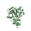
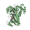
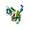
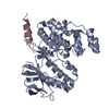
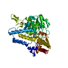
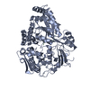
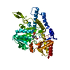
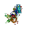
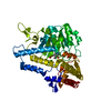
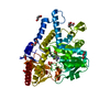
A: Forkhead box protein M1
B: Forkhead box protein M1
C: DNA (5'-D(P*AP*AP*AP*TP*TP*GP*TP*TP*TP*AP*TP*AP*AP*AP*CP*AP*GP*CP*CP*CP*G)-3')
D: DNA (5'-D(P*TP*TP*CP*GP*GP*GP*CP*TP*GP*TP*TP*TP*AP*TP*AP*AP*AP*CP*AP*AP*T)-3')
hetero molecules


Theoretical massNumber of molelcules
Total (without water)46,1336
Polymers46,0844
Non-polymers492
Water2,936163
1


  • Idetical with deposited unit
  • defined by author&software
TypeNameSymmetry operationNumber
identity operation1_555x,y,z1
Buried area5270 Å2
ΔGint-64 kcal/mol
Surface area16980 Å2
MethodPISA
Unit cell
Length a, b, c (Å)63.070, 119.750, 152.970
Angle α, β, γ (deg.)90.00, 90.00, 90.00
Int Tables number20
Space group name H-MC2221

-
Components

#1: Protein Forkhead box protein M1 / Forkhead-related protein FKHL16 / Hepatocyte nuclear factor 3 forkhead homolog 11 / HNF-3/fork-head ...Forkhead-related protein FKHL16 / Hepatocyte nuclear factor 3 forkhead homolog 11 / HNF-3/fork-head homolog 11 / HFH-11 / Winged-helix factor from INS-1 cells / M-phase phosphoprotein 2 / MPM-2 reactive phosphoprotein 2 / Transcription factor Trident


Mass: 16600.861 Da / Num. of mol.: 2 / Fragment: DNA-binding domain, UNP residues 222-360
Source method: isolated from a genetically manipulated source
Source: (gene. exp.) Homo sapiens (human) / Gene: FKHL16, FOXM1, HFH11, MPP2, WIN / Plasmid: pET28-LIC / Production host: Escherichia coli (E. coli) / Strain (production host): BL21 (DE3) / References: UniProt: Q08050
#2: DNA chain DNA (5'-D(P*AP*AP*AP*TP*TP*GP*TP*TP*TP*AP*TP*AP*AP*AP*CP*AP*GP*CP*CP*CP*G)-3')


Mass: 6430.198 Da / Num. of mol.: 1 / Source method: obtained synthetically / Details: DNA oligo
#3: DNA chain DNA (5'-D(P*TP*TP*CP*GP*GP*GP*CP*TP*GP*TP*TP*TP*AP*TP*AP*AP*AP*CP*AP*AP*T)-3')


Mass: 6452.195 Da / Num. of mol.: 1 / Source method: obtained synthetically / Details: DNA oligo
#4: Chemical ChemComp-MG / MAGNESIUM ION


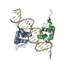
Mass: 24.305 Da / Num. of mol.: 2 / Source method: obtained synthetically / Formula: Mg
#5: Water ChemComp-HOH / water


Mass: 18.015 Da / Num. of mol.: 163 / Source method: isolated from a natural source / Formula: H2O

-
Experimental details

-
Experiment

ExperimentMethod: X-RAY DIFFRACTION / Number of used crystals: 1

-
Sample preparation

CrystalDensity Matthews: 3.1 Å3/Da / Density % sol: 60.38 %
Crystal growTemperature: 291 K / Method: vapor diffusion, hanging drop / pH: 7.5
Details: 24% PEG3350, 0.2M Sodium Malonate, pH7.5, 3uL Protein at 12g/L mixed with DNA at 0.5mM added tO 3uL of reservoir, VAPOR DIFFUSION, HANGING DROP, temperature 291K
Components of the solutions
IDNameCrystal-IDSol-ID
1PEG335011
2Sodium Malonate11
3HOH11
4PEG335012
5Sodium Malonate12
6HOH12

-
Data collection

DiffractionMean temperature: 100 K
Diffraction sourceSource: SYNCHROTRON / Site: ESRF / Beamline: ID23-1 / Wavelength: 1.0723 / Wavelength: 1.0723 Å
DetectorType: ADSC QUANTUM Q315r / Detector: CCD / Date: Nov 1, 2008
RadiationProtocol: SINGLE WAVELENGTH / Monochromatic (M) / Laue (L): M / Scattering type: x-ray
Radiation wavelengthWavelength: 1.0723 Å / Relative weight: 1
ReflectionResolution: 2.2→76 Å / Num. obs: 27165 / % possible obs: 91.4 % / Observed criterion σ(I): 0 / Redundancy: 5 % / Biso Wilson estimate: 48.1 Å2 / Rmerge(I) obs: 0.058 / Net I/σ(I): 16.1
Reflection shellResolution: 2.2→2.32 Å / Redundancy: 4.4 % / Rmerge(I) obs: 0.45 / Mean I/σ(I) obs: 3.1 / % possible all: 59.4

-
Processing

Software
NameVersionClassification
DNAdata collection
AMoREphasing
REFMAC5.5.0063refinement
MOSFLMdata reduction
SCALAdata scaling
RefinementMethod to determine structure: MOLECULAR REPLACEMENT
Starting model: PDB ENTRY 2C6Y
Resolution: 2.21→32.94 Å / Cor.coef. Fo:Fc: 0.95 / Cor.coef. Fo:Fc free: 0.939 / SU B: 9.275 / SU ML: 0.105 / TLS residual ADP flag: LIKELY RESIDUAL / Cross valid method: THROUGHOUT / ESU R: 0.195 / ESU R Free: 0.173 / Stereochemistry target values: MAXIMUM LIKELIHOOD / Details: HYDROGENS HAVE BEEN ADDED IN THE RIDING POSITIONS
RfactorNum. reflection% reflectionSelection details
Rfree0.23425 1389 5.2 %RANDOM
Rwork0.204 ---
obs0.20557 25545 91.44 %-
Solvent computationIon probe radii: 0.8 Å / Shrinkage radii: 0.8 Å / VDW probe radii: 1.2 Å / Solvent model: MASK
Displacement parametersBiso mean: 19.23 Å2
Baniso -1Baniso -2Baniso -3
1-1.22 Å20 Å20 Å2
2--0.23 Å20 Å2
3----1.45 Å2
Refinement stepCycle: LAST / Resolution: 2.21→32.94 Å
ProteinNucleic acidLigandSolventTotal
Num. atoms1560 861 2 163 2586
Refine LS restraints
Refine-IDTypeDev idealDev ideal targetNumber
X-RAY DIFFRACTIONr_bond_refined_d0.0090.0212582
X-RAY DIFFRACTIONr_bond_other_d0.0010.021510
X-RAY DIFFRACTIONr_angle_refined_deg1.4462.3543676
X-RAY DIFFRACTIONr_angle_other_deg0.92233694
X-RAY DIFFRACTIONr_dihedral_angle_1_deg5.8315185
X-RAY DIFFRACTIONr_dihedral_angle_2_deg31.39522.37580
X-RAY DIFFRACTIONr_dihedral_angle_3_deg13.63915276
X-RAY DIFFRACTIONr_dihedral_angle_4_deg15.8121512
X-RAY DIFFRACTIONr_chiral_restr0.0660.2393
X-RAY DIFFRACTIONr_gen_planes_refined0.0050.0212201
X-RAY DIFFRACTIONr_gen_planes_other0.0010.02450
X-RAY DIFFRACTIONr_nbd_refined
X-RAY DIFFRACTIONr_nbd_other
X-RAY DIFFRACTIONr_nbtor_refined
X-RAY DIFFRACTIONr_nbtor_other
X-RAY DIFFRACTIONr_xyhbond_nbd_refined
X-RAY DIFFRACTIONr_xyhbond_nbd_other
X-RAY DIFFRACTIONr_metal_ion_refined
X-RAY DIFFRACTIONr_metal_ion_other
X-RAY DIFFRACTIONr_symmetry_vdw_refined
X-RAY DIFFRACTIONr_symmetry_vdw_other
X-RAY DIFFRACTIONr_symmetry_hbond_refined
X-RAY DIFFRACTIONr_symmetry_hbond_other
X-RAY DIFFRACTIONr_symmetry_metal_ion_refined
X-RAY DIFFRACTIONr_symmetry_metal_ion_other
X-RAY DIFFRACTIONr_mcbond_it1.0592933
X-RAY DIFFRACTIONr_mcbond_other0.2112362
X-RAY DIFFRACTIONr_mcangle_it1.78231523
X-RAY DIFFRACTIONr_scbond_it0.96521649
X-RAY DIFFRACTIONr_scangle_it1.54532152
X-RAY DIFFRACTIONr_rigid_bond_restr
X-RAY DIFFRACTIONr_sphericity_free
X-RAY DIFFRACTIONr_sphericity_bonded
LS refinement shellResolution: 2.21→2.267 Å / Total num. of bins used: 20
RfactorNum. reflection% reflection
Rfree0.409 69 -
Rwork0.331 1117 -
obs--54.88 %
Refinement TLS params.

Method: refined / Refine-ID: X-RAY DIFFRACTION

IDL112)L122)L132)L222)L232)L332)S11 (Å °)S12 (Å °)S13 (Å °)S21 (Å °)S22 (Å °)S23 (Å °)S31 (Å °)S32 (Å °)S33 (Å °)T112)T122)T132)T222)T232)T332)Origin x (Å)Origin y (Å)Origin z (Å)
13.8974-1.9459-0.75144.93820.92014.1264-0.1242-0.0764-0.09770.35350.1006-0.30560.17590.20090.02370.02930.0188-0.01870.14330.0410.049210.297429.182212.9834
28.95711.7863-0.33564.39130.55347.6112-0.34970.8179-0.3373-0.25950.14240.05230.1524-0.0090.20730.04360.01020.02260.1841-0.01880.041410.262523.7499-2.3788
33.48030.0694-0.76813.8758-1.74329.72470.22540.0460.09790.26330.07840.277-0.1218-0.9885-0.30380.05550.05570.0220.21980.01970.0253-4.467136.216325.5536
Refinement TLS group
IDRefine-IDRefine TLS-IDAuth asym-IDAuth seq-ID
1X-RAY DIFFRACTION1C1 - 21
2X-RAY DIFFRACTION1D1 - 21
3X-RAY DIFFRACTION2A232 - 321
4X-RAY DIFFRACTION3B235 - 327

+
About Yorodumi

-
News

-
Feb 9, 2022. New format data for meta-information of EMDB entries

New format data for meta-information of EMDB entries

  • Version 3 of the EMDB header file is now the official format.
  • The previous official version 1.9 will be removed from the archive.

Related info.:EMDB header

External links:wwPDB to switch to version 3 of the EMDB data model

-
Aug 12, 2020. Covid-19 info

Covid-19 info

URL: https://pdbjlvh1.pdbj.org/emnavi/covid19.php

New page: Covid-19 featured information page in EM Navigator.

Related info.:Covid-19 info / Mar 5, 2020. Novel coronavirus structure data

+
Mar 5, 2020. Novel coronavirus structure data

Novel coronavirus structure data

Related info.:Yorodumi Speices / Aug 12, 2020. Covid-19 info

External links:COVID-19 featured content - PDBj / Molecule of the Month (242):Coronavirus Proteases

+
Jan 31, 2019. EMDB accession codes are about to change! (news from PDBe EMDB page)

EMDB accession codes are about to change! (news from PDBe EMDB page)

  • The allocation of 4 digits for EMDB accession codes will soon come to an end. Whilst these codes will remain in use, new EMDB accession codes will include an additional digit and will expand incrementally as the available range of codes is exhausted. The current 4-digit format prefixed with “EMD-” (i.e. EMD-XXXX) will advance to a 5-digit format (i.e. EMD-XXXXX), and so on. It is currently estimated that the 4-digit codes will be depleted around Spring 2019, at which point the 5-digit format will come into force.
  • The EM Navigator/Yorodumi systems omit the EMD- prefix.

Related info.:Q: What is EMD? / ID/Accession-code notation in Yorodumi/EM Navigator

External links:EMDB Accession Codes are Changing Soon! / Contact to PDBj

+
Jul 12, 2017. Major update of PDB

Major update of PDB

  • wwPDB released updated PDB data conforming to the new PDBx/mmCIF dictionary.
  • This is a major update changing the version number from 4 to 5, and with Remediation, in which all the entries are updated.
  • In this update, many items about electron microscopy experimental information are reorganized (e.g. em_software).
  • Now, EM Navigator and Yorodumi are based on the updated data.

External links:wwPDB Remediation / Enriched Model Files Conforming to OneDep Data Standards Now Available in the PDB FTP Archive

-
Yorodumi

Thousand views of thousand structures

  • Yorodumi is a browser for structure data from EMDB, PDB, SASBDB, etc.
  • This page is also the successor to EM Navigator detail page, and also detail information page/front-end page for Omokage search.
  • The word "yorodu" (or yorozu) is an old Japanese word meaning "ten thousand". "mi" (miru) is to see.

Related info.:EMDB / PDB / SASBDB / Comparison of 3 databanks / Yorodumi Search / Aug 31, 2016. New EM Navigator & Yorodumi / Yorodumi Papers / Jmol/JSmol / Function and homology information / Changes in new EM Navigator and Yorodumi

Read more