 Movie
Movie Controller

[English] 日本語
 Yorodumi
- PDB-3g4d: Crystal Structure of (+)-delta-Cadinene Synthase from Gossypium a... -
+ Open data

- Basic information
| Entry | Database: PDB / ID: 3g4d | ||||||
|---|---|---|---|---|---|---|---|
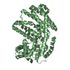
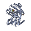
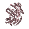
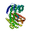
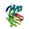
| Title | Crystal Structure of (+)-delta-Cadinene Synthase from Gossypium arboreum and Evolutionary Divergence of Metal Binding Motifs for Catalysis | ||||||
 Components Components | (+)-delta-cadinene synthase isozyme XC1 | ||||||
 Keywords Keywords | LYASE / cyclase / Magnesium / Metal-binding | ||||||
| Function / homology |  Function and homology information Function and homology information(+)-delta-cadinene synthase / (+)-delta-cadinene synthase activity / diterpenoid biosynthetic process / magnesium ion binding Similarity search - Function | ||||||
| Biological species |  | ||||||
| Method |  X-RAY DIFFRACTION / SYNCHROTRON / MOLECULAR REPLACEMENT / Resolution: 2.403 Å X-RAY DIFFRACTION / SYNCHROTRON / MOLECULAR REPLACEMENT / Resolution: 2.403 Å | ||||||
 Authors Authors | Gennadios, H.A. / Di Costanzo, L. / Miller, D.J. / Allemann, R.K. / Christianson, D.W. | ||||||
 Citation Citation | Journal: Biochemistry / Year: 2009 Title: Crystal structure of (+)-delta-cadinene synthase from Gossypium arboreum and evolutionary divergence of metal binding motifs for catalysis. Authors: Gennadios, H.A. / Gonzalez, V. / Di Costanzo, L. / Li, A. / Yu, F. / Miller, D.J. / Allemann, R.K. / Christianson, D.W. | ||||||
| History |
|
- Structure visualization
| Structure viewer | Molecule: MolmilJmol/JSmol |
|---|
- Downloads & links
-Download
| PDBx/mmCIF format | 3g4d.cif.gz | 227.9 KB | Display | PDBx/mmCIF format |
|---|---|---|---|---|
| PDB format | pdb3g4d.ent.gz | 182.5 KB | Display | PDB format |
| PDBx/mmJSON format | 3g4d.json.gz | Tree view | PDBx/mmJSON format | |
| Others |  Other downloads Other downloads |
-Validation report
| Summary document | 3g4d_validation.pdf.gz | 467.7 KB | Display | wwPDB validaton report |
|---|---|---|---|---|
| Full document | 3g4d_full_validation.pdf.gz | 498.6 KB | Display | |
| Data in XML | 3g4d_validation.xml.gz | 43.8 KB | Display | |
| Data in CIF | 3g4d_validation.cif.gz | 61.6 KB | Display | |
| Arichive directory | https://data.pdbj.org/pub/pdb/validation_reports/g4/3g4dftp://data.pdbj.org/pub/pdb/validation_reports/g4/3g4d | HTTPS FTP |
-Related structure data
| Related structure data |  3g4fC  5eatS C: citing same article ( S: Starting model for refinement |
|---|---|
| Similar structure data |


-Links
 PDBj
PDBj


- Assembly
| Deposited unit | 
| ||||||||
|---|---|---|---|---|---|---|---|---|---|
| 1 | 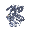
| ||||||||
| 2 | 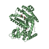
| ||||||||
| Unit cell |
|
-Components
| #1: Protein | Mass: 64217.648 Da / Num. of mol.: 2 Source method: isolated from a genetically manipulated source Source: (gene. exp.)  #2: Chemical |   Mass: 78.133 Da / Num. of mol.: 3 / Source method: obtained synthetically / Formula: C2H6OS Mass: 78.133 Da / Num. of mol.: 3 / Source method: obtained synthetically / Formula: C2H6OS#3: Chemical | ChemComp-GOL /   Mass: 92.094 Da / Num. of mol.: 4 / Source method: obtained synthetically / Formula: C3H8O3 Mass: 92.094 Da / Num. of mol.: 4 / Source method: obtained synthetically / Formula: C3H8O3#4: Water | ChemComp-HOH / |  Mass: 18.015 Da / Num. of mol.: 382 / Source method: isolated from a natural source / Formula: H2O Mass: 18.015 Da / Num. of mol.: 382 / Source method: isolated from a natural source / Formula: H2O |
|---|
-Experimental details
-Experiment
| Experiment | Method: X-RAY DIFFRACTION / Number of used crystals: 1 |
|---|
- Sample preparation
| Crystal | Density Matthews: 2.57 Å3/Da / Density % sol: 52.12 % |
|---|---|
| Crystal grow | Temperature: 298 K / Method: vapor diffusion, hanging drop / pH: 8 Details: drop containing 5 L protein solution (10 mg/mL DCS, 20 mM Tris (pH 8.0), 2 mM MgCl2, 5 mM BME) and 5 L precipitant solution (100 mM Tris (pH 7.5), 200 mM Li2SO4, 15-17% polyethylene glycol ...Details: drop containing 5 L protein solution (10 mg/mL DCS, 20 mM Tris (pH 8.0), 2 mM MgCl2, 5 mM BME) and 5 L precipitant solution (100 mM Tris (pH 7.5), 200 mM Li2SO4, 15-17% polyethylene glycol (PEG) 4000, 100 mM BaCl2 2H2O) was suspended over a reservoir of 600 L precipitant solution. , VAPOR DIFFUSION, HANGING DROP, temperature 298K |
-Data collection
| Diffraction | Mean temperature: 100 K |
|---|---|
| Diffraction source | Source: SYNCHROTRON / Site: APS  / Beamline: 24-ID-C / Beamline: 24-ID-C |
| Detector | Type: ADSC QUANTUM 315r / Detector: CCD |
| Radiation | Protocol: SINGLE WAVELENGTH / Monochromatic (M) / Laue (L): M / Scattering type: x-ray |
| Radiation wavelength | Relative weight: 1 |
| Reflection | Resolution: 2.4→50 Å / Num. all: 51262 / Num. obs: 51262 / % possible obs: 99.4 % / Redundancy: 6.1 % / Rmerge(I) obs: 0.092 / Rsym value: 0.092 / Net I/σ(I): 19.7 |
| Reflection shell | Resolution: 2.4→2.49 Å / Redundancy: 6.2 % / Rmerge(I) obs: 0.525 / Mean I/σ(I) obs: 4.65 / Num. unique all: 5144 / Rsym value: 0.525 |
- Processing
| Software |
| ||||||||||||||||||||||||||||||||||||||||||||||||||||||||||||||||||||||||||||||||||||||||||||||||||||||||||||||||
|---|---|---|---|---|---|---|---|---|---|---|---|---|---|---|---|---|---|---|---|---|---|---|---|---|---|---|---|---|---|---|---|---|---|---|---|---|---|---|---|---|---|---|---|---|---|---|---|---|---|---|---|---|---|---|---|---|---|---|---|---|---|---|---|---|---|---|---|---|---|---|---|---|---|---|---|---|---|---|---|---|---|---|---|---|---|---|---|---|---|---|---|---|---|---|---|---|---|---|---|---|---|---|---|---|---|---|---|---|---|---|---|---|---|
| Refinement | Method to determine structure: MOLECULAR REPLACEMENT Starting model: Nicotiana tabacum 5-epi aristolochene synthase (PDB ENTRY 5EAT) Resolution: 2.403→47.698 Å / SU ML: 0.36 / σ(F): 1.34 / Stereochemistry target values: ML
| ||||||||||||||||||||||||||||||||||||||||||||||||||||||||||||||||||||||||||||||||||||||||||||||||||||||||||||||||
| Solvent computation | Shrinkage radii: 0.9 Å / VDW probe radii: 1.11 Å / Solvent model: FLAT BULK SOLVENT MODEL / Bsol: 71.95 Å2 / ksol: 0.345 e/Å3 | ||||||||||||||||||||||||||||||||||||||||||||||||||||||||||||||||||||||||||||||||||||||||||||||||||||||||||||||||
| Refinement step | Cycle: LAST / Resolution: 2.403→47.698 Å
| ||||||||||||||||||||||||||||||||||||||||||||||||||||||||||||||||||||||||||||||||||||||||||||||||||||||||||||||||
| Refine LS restraints |
| ||||||||||||||||||||||||||||||||||||||||||||||||||||||||||||||||||||||||||||||||||||||||||||||||||||||||||||||||
| LS refinement shell |
|
