Resolution: 2.02→2.05 Å / Redundancy: 2.7 % / Rmerge(I) obs: 0.524 / Mean I/σ(I) obs: 2 / Num. unique all: 1793 / Χ2: 0.967 / % possible all: 92.1
-
Processing
Software
Name
Version
Classification
NB
DENZO
datareduction
SCALEPACK
datascaling
PHENIX
refinement
PDB_EXTRACT
3.006
dataextraction
SBC-Collect
datacollection
HKL-3000
datareduction
PHENIX
phasing
Refinement
Method to determine structure: SAD / Resolution: 2.009→26.995 Å / Occupancy max: 1 / Occupancy min: 0 / FOM work R set: 0.834 / σ(F): 1.89 / σ(I): 0 / Stereochemistry target values: TWIN_LSQ_F Details: Refinement was performed using twin law -h,-k,l and twin fraction 0.49. Unknown ligands were modeled as waters in chains X and Y. THE FRIEDEL PAIRS WERE USED FOR PHASING.
Rfactor
Num. reflection
% reflection
Rfree
0.223
3810
5.33 %
Rwork
0.181
-
-
all
0.183
71468
-
obs
0.183
71468
93.54 %
Solvent computation
Shrinkage radii: 0.9 Å / VDW probe radii: 1.11 Å / Solvent model: FLAT BULK SOLVENT MODEL / Bsol: 52.195 Å2 / ksol: 0.363 e/Å3
In the structure databanks used in Yorodumi, some data are registered as the other names, "COVID-19 virus" and "2019-nCoV". Here are the details of the virus and the list of structure data.
Jan 31, 2019. EMDB accession codes are about to change! (news from PDBe EMDB page)
EMDB accession codes are about to change! (news from PDBe EMDB page)
The allocation of 4 digits for EMDB accession codes will soon come to an end. Whilst these codes will remain in use, new EMDB accession codes will include an additional digit and will expand incrementally as the available range of codes is exhausted. The current 4-digit format prefixed with “EMD-” (i.e. EMD-XXXX) will advance to a 5-digit format (i.e. EMD-XXXXX), and so on. It is currently estimated that the 4-digit codes will be depleted around Spring 2019, at which point the 5-digit format will come into force.
The EM Navigator/Yorodumi systems omit the EMD- prefix.
Related info.:Q: What is EMD? / ID/Accession-code notation in Yorodumi/EM Navigator
Yorodumi is a browser for structure data from EMDB, PDB, SASBDB, etc.
This page is also the successor to EM Navigator detail page, and also detail information page/front-end page for Omokage search.
The word "yorodu" (or yorozu) is an old Japanese word meaning "ten thousand". "mi" (miru) is to see.
Related info.:EMDB / PDB / SASBDB / Comparison of 3 databanks / Yorodumi Search / Aug 31, 2016. New EM Navigator & Yorodumi / Yorodumi Papers / Jmol/JSmol / Function and homology information / Changes in new EM Navigator and Yorodumi
 Movie
Movie Controller

 Yorodumi
 Open data
 Basic information
 Components
 Keywords
 Function and homology information
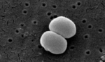 Staphylococcus epidermidis (bacteria)
 X-RAY DIFFRACTION /
  Authors
 Citation
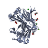
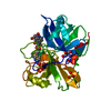
 Structure visualization
 Downloads & links
 Other downloads
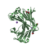


 PDBj
PDBj Assembly
 Mass: 18.015 Da / Num. of mol.: 448 / Source method: isolated from a natural source / Formula: H2O
 Sample preparation
 / Beamline: 19-BM / Wavelength: 0.9792 Å
 Processing