Type: MAR CCD 165 mm / Detector: CCD / Date: Nov 29, 2007
Radiation
Protocol: SINGLE WAVELENGTH / Monochromatic (M) / Laue (L): M / Scattering type: x-ray
Radiation wavelength
Wavelength: 0.979 Å / Relative weight: 1
Reflection
Resolution: 1.9→30 Å / Num. obs: 41038 / % possible obs: 98.4 % / Observed criterion σ(I): -3 / Redundancy: 6 % / Rmerge(I) obs: 0.092 / Net I/σ(I): 21.1
Reflection shell
Resolution: 1.9→1.97 Å / Redundancy: 4.6 % / Rmerge(I) obs: 0.299 / Mean I/σ(I) obs: 3.3 / % possible all: 92.2
-
Processing
Software
Name
Version
Classification
ADSC
Quantum
datacollection
SnB
phasing
REFMAC
5.2.0019
refinement
HKL-2000
datareduction
SCALEPACK
datascaling
Refinement
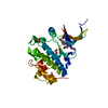
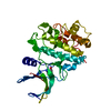
Method to determine structure: SAD / Resolution: 1.9→22.43 Å / Cor.coef. Fo:Fc: 0.915 / Cor.coef. Fo:Fc free: 0.909 / SU B: 8.109 / SU ML: 0.11 / TLS residual ADP flag: LIKELY RESIDUAL / Cross valid method: THROUGHOUT / ESU R: 0.198 / ESU R Free: 0.167 / Stereochemistry target values: MAXIMUM LIKELIHOOD Details: Authors explanation on why Rfree/R are high at this resolution: The analyses of the Patterson function reveals a significant off-origin peak that is 56.98 % of the origin peak, indicating ...Details: Authors explanation on why Rfree/R are high at this resolution: The analyses of the Patterson function reveals a significant off-origin peak that is 56.98 % of the origin peak, indicating pseudo translational symmetry. The chance of finding a peak of this or larger height by random in a structure without pseudo translational symmetry is equal to the 2.5284e-05. The detected tranlational NCS is most likely also responsible for the elevated intensity ratio. The results of the L-test indicate that the intensity statistics behave as expected. No twinning is suspected.
Rfactor
Num. reflection
% reflection
Selection details
Rfree
0.28875
1006
5.2 %
RANDOM
Rwork
0.27544
-
-
-
obs
0.27613
18375
100 %
-
Solvent computation
Ion probe radii: 0.8 Å / Shrinkage radii: 0.8 Å / VDW probe radii: 1.2 Å / Solvent model: BABINET MODEL WITH MASK
Displacement parameters
Biso mean: 23.793 Å2
Baniso -1
Baniso -2
Baniso -3
1-
-0.05 Å2
0 Å2
0 Å2
2-
-
0.1 Å2
0 Å2
3-
-
-
-0.05 Å2
Refinement step
Cycle: LAST / Resolution: 1.9→22.43 Å
Protein
Nucleic acid
Ligand
Solvent
Total
Num. atoms
1494
0
10
121
1625
Refine LS restraints
Refine-ID
Type
Dev ideal
Dev ideal target
Number
X-RAY DIFFRACTION
r_bond_refined_d
0.009
0.022
1529
X-RAY DIFFRACTION
r_bond_other_d
X-RAY DIFFRACTION
r_angle_refined_deg
1.258
2.014
2064
X-RAY DIFFRACTION
r_angle_other_deg
X-RAY DIFFRACTION
r_dihedral_angle_1_deg
6.789
5
202
X-RAY DIFFRACTION
r_dihedral_angle_2_deg
29.552
22.857
56
X-RAY DIFFRACTION
r_dihedral_angle_3_deg
14.707
15
285
X-RAY DIFFRACTION
r_dihedral_angle_4_deg
14.98
15
16
X-RAY DIFFRACTION
r_chiral_restr
0.071
0.2
258
X-RAY DIFFRACTION
r_gen_planes_refined
0.004
0.02
1086
X-RAY DIFFRACTION
r_gen_planes_other
X-RAY DIFFRACTION
r_nbd_refined
0.23
0.2
629
X-RAY DIFFRACTION
r_nbd_other
X-RAY DIFFRACTION
r_nbtor_refined
0.294
0.2
1029
X-RAY DIFFRACTION
r_nbtor_other
X-RAY DIFFRACTION
r_xyhbond_nbd_refined
0.142
0.2
105
X-RAY DIFFRACTION
r_xyhbond_nbd_other
X-RAY DIFFRACTION
r_metal_ion_refined
X-RAY DIFFRACTION
r_metal_ion_other
X-RAY DIFFRACTION
r_symmetry_vdw_refined
0.276
0.2
78
X-RAY DIFFRACTION
r_symmetry_vdw_other
X-RAY DIFFRACTION
r_symmetry_hbond_refined
0.216
0.2
11
X-RAY DIFFRACTION
r_symmetry_hbond_other
X-RAY DIFFRACTION
r_symmetry_metal_ion_refined
0.423
0.2
1
X-RAY DIFFRACTION
r_symmetry_metal_ion_other
X-RAY DIFFRACTION
r_mcbond_it
0.571
1.5
997
X-RAY DIFFRACTION
r_mcbond_other
X-RAY DIFFRACTION
r_mcangle_it
1.125
2
1589
X-RAY DIFFRACTION
r_scbond_it
2.332
3
532
X-RAY DIFFRACTION
r_scangle_it
3.721
4.5
472
X-RAY DIFFRACTION
r_rigid_bond_restr
X-RAY DIFFRACTION
r_sphericity_free
X-RAY DIFFRACTION
r_sphericity_bonded
LS refinement shell
Resolution: 1.9→1.949 Å / Total num. of bins used: 20
Rfactor
Num. reflection
% reflection
Rfree
0.292
59
-
Rwork
0.267
1042
-
obs
-
-
100 %
Refinement TLS params.
Method: refined / Origin x: 31.1109 Å / Origin y: 14.653 Å / Origin z: 18.9432 Å
11
12
13
21
22
23
31
32
33
T
-0.0723 Å2
0.0017 Å2
0.0056 Å2
-
-0.1525 Å2
-0.0018 Å2
-
-
-0.0343 Å2
L
1.4752 °2
-0.0024 °2
0.2561 °2
-
0.5986 °2
-0.0285 °2
-
-
3.4684 °2
S
0.0563 Å °
0.0042 Å °
0.1196 Å °
0.0077 Å °
-0.0172 Å °
0.0001 Å °
-0.132 Å °
0.006 Å °
-0.0391 Å °
Refinement TLS group
ID
Refine-ID
Refine TLS-ID
Auth asym-ID
Auth seq-ID
1
X-RAY DIFFRACTION
1
B
118 - 182
2
X-RAY DIFFRACTION
1
A
118 - 175
3
X-RAY DIFFRACTION
1
B
116 - 117
4
X-RAY DIFFRACTION
1
A
116 - 117
5
X-RAY DIFFRACTION
1
B
1 - 102
6
X-RAY DIFFRACTION
1
A
1 - 102
+
About Yorodumi
-
News
-
Feb 9, 2022. New format data for meta-information of EMDB entries
New format data for meta-information of EMDB entries
Version 3 of the EMDB header file is now the official format.
The previous official version 1.9 will be removed from the archive.
In the structure databanks used in Yorodumi, some data are registered as the other names, "COVID-19 virus" and "2019-nCoV". Here are the details of the virus and the list of structure data.
Jan 31, 2019. EMDB accession codes are about to change! (news from PDBe EMDB page)
EMDB accession codes are about to change! (news from PDBe EMDB page)
The allocation of 4 digits for EMDB accession codes will soon come to an end. Whilst these codes will remain in use, new EMDB accession codes will include an additional digit and will expand incrementally as the available range of codes is exhausted. The current 4-digit format prefixed with “EMD-” (i.e. EMD-XXXX) will advance to a 5-digit format (i.e. EMD-XXXXX), and so on. It is currently estimated that the 4-digit codes will be depleted around Spring 2019, at which point the 5-digit format will come into force.
The EM Navigator/Yorodumi systems omit the EMD- prefix.
Related info.:Q: What is EMD? / ID/Accession-code notation in Yorodumi/EM Navigator
Yorodumi is a browser for structure data from EMDB, PDB, SASBDB, etc.
This page is also the successor to EM Navigator detail page, and also detail information page/front-end page for Omokage search.
The word "yorodu" (or yorozu) is an old Japanese word meaning "ten thousand". "mi" (miru) is to see.
Related info.:EMDB / PDB / SASBDB / Comparison of 3 databanks / Yorodumi Search / Aug 31, 2016. New EM Navigator & Yorodumi / Yorodumi Papers / Jmol/JSmol / Function and homology information / Changes in new EM Navigator and Yorodumi
 Movie
Movie Controller

 Yorodumi
 Open data
 Basic information
 Components
 Keywords
 Function and homology information
 Bordetella parapertussis (bacteria)
 X-RAY DIFFRACTION /
  Authors
 Citation
 Structure visualization
 Downloads & links
 Other downloads


 PDBj
PDBj Assembly

 Mass: 22.990 Da / Num. of mol.: 3 / Source method: obtained synthetically / Formula: Na
 Mass: 106.120 Da / Num. of mol.: 1 / Source method: obtained synthetically / Formula: C4H10O3
 Mass: 18.015 Da / Num. of mol.: 121 / Source method: isolated from a natural source / Formula: H2O
 Sample preparation
 / Beamline: X4C / Wavelength: 0.979 Å
 Processing