 Movie
Movie Controller

[English] 日本語
 Yorodumi
- PDB-3fbq: The crystal structure of the conserved domain protein from Bacill... -
+ Open data

- Basic information
| Entry | Database: PDB / ID: 3fbq | ||||||
|---|---|---|---|---|---|---|---|
| Title | The crystal structure of the conserved domain protein from Bacillus anthracis | ||||||
 Components Components | Conserved domain protein | ||||||
 Keywords Keywords | structural genomics / unknown function / conserved domain protein / Bacillus anthracis / PSI2 / MCSG / Protein Structure Initiative / Midwest Center for Structural Genomics | ||||||
| Function / homology |  Function and homology information Function and homology informationbacillus anthracis domain / Conserved domain protein. / Domain of unknown function DUF4179 / Domain of unknown function DUF5643 / Domain of unknown function (DUF4179) / Family of unknown function (DUF5643) / Immunoglobulin-like / Sandwich / Mainly Beta Similarity search - Domain/homology | ||||||
| Biological species |  | ||||||
| Method |  X-RAY DIFFRACTION / SYNCHROTRON / MAD / Resolution: 2.71 Å X-RAY DIFFRACTION / SYNCHROTRON / MAD / Resolution: 2.71 Å | ||||||
 Authors Authors | Zhang, R. / Joachimiak, G. / Kim, Y. / Gornicki, P. / Joachimiak, A. / Midwest Center for Structural Genomics (MCSG) | ||||||
 Citation Citation | Journal: To be Published Title: The crystal structure of the conserved domain protein from Bacillus anthracis Authors: Zhang, R. / Joachimiak, G. / Kim, Y. / Gornicki, P. / Joachimiak, A. | ||||||
| History |
|
- Structure visualization
| Structure viewer | Molecule: MolmilJmol/JSmol |
|---|
- Downloads & links
-Download
| PDBx/mmCIF format | 3fbq.cif.gz | 67.6 KB | Display | PDBx/mmCIF format |
|---|---|---|---|---|
| PDB format | pdb3fbq.ent.gz | 51.3 KB | Display | PDB format |
| PDBx/mmJSON format | 3fbq.json.gz | Tree view | PDBx/mmJSON format | |
| Others |  Other downloads Other downloads |
-Validation report
| Arichive directory | https://data.pdbj.org/pub/pdb/validation_reports/fb/3fbqftp://data.pdbj.org/pub/pdb/validation_reports/fb/3fbq | HTTPS FTP |
|---|
-Related structure data
| Similar structure data | |
|---|---|
| Other databases |


-Links
 PDBj
PDBj- Assembly
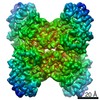
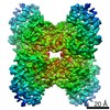
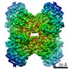
| Deposited unit | 
| ||||||||
|---|---|---|---|---|---|---|---|---|---|
| 1 | 
| ||||||||
| Unit cell |
| ||||||||
| Details | This protein existed as trimer. The second and third part of the biological are genarated by the three fold axis: z,x,y and y,z,x |
-Components
| #1: Protein | Mass: 32591.557 Da / Num. of mol.: 1 Source method: isolated from a genetically manipulated source Source: (gene. exp.) |
|---|---|
| #2: Water | ChemComp-HOH /  Mass: 18.015 Da / Num. of mol.: 27 / Source method: isolated from a natural source / Formula: H2O Mass: 18.015 Da / Num. of mol.: 27 / Source method: isolated from a natural source / Formula: H2O |
-Experimental details
-Experiment
| Experiment | Method: X-RAY DIFFRACTION / Number of used crystals: 1 |
|---|
- Sample preparation
| Crystal | Density Matthews: 2.67 Å3/Da / Density % sol: 53.87 % |
|---|---|
| Crystal grow | Temperature: 289 K / Method: vapor diffusion, sitting drop / pH: 7 Details: 2M (NH4)2SO4, 0.1M Tris and 0.2M Li2SO4, pH 7, VAPOR DIFFUSION, SITTING DROP, temperature 289K |
-Data collection
| Diffraction | Mean temperature: 100 K | |||||||||
|---|---|---|---|---|---|---|---|---|---|---|
| Diffraction source | Source: SYNCHROTRON / Site: APS  / Beamline: 19-ID / Wavelength: 0.9794, 0.9796 / Beamline: 19-ID / Wavelength: 0.9794, 0.9796 | |||||||||
| Detector | Type: ADSC QUANTUM 315 / Detector: CCD / Date: Dec 3, 2007 / Details: mirrors | |||||||||
| Radiation | Monochromator: Si 111 channel / Protocol: MAD / Monochromatic (M) / Laue (L): M / Scattering type: x-ray | |||||||||
| Radiation wavelength |
| |||||||||
| Reflection | Resolution: 2.7→71.8 Å / Num. all: 9267 / Num. obs: 9226 / % possible obs: 99.56 % / Observed criterion σ(F): 2 / Observed criterion σ(I): 2 / Redundancy: 8.5 % / Biso Wilson estimate: 52 Å2 / Rmerge(I) obs: 0.127 / Net I/σ(I): 21.45 | |||||||||
| Reflection shell | Resolution: 2.7→2.77 Å / Redundancy: 8.5 % / Rmerge(I) obs: 0.795 / Mean I/σ(I) obs: 2.72 / Num. unique all: 696 / % possible all: 97.41 |
- Processing
| Software |
| |||||||||||||||||||||||||||||||||||||||||||||||||||||||||||||||||
|---|---|---|---|---|---|---|---|---|---|---|---|---|---|---|---|---|---|---|---|---|---|---|---|---|---|---|---|---|---|---|---|---|---|---|---|---|---|---|---|---|---|---|---|---|---|---|---|---|---|---|---|---|---|---|---|---|---|---|---|---|---|---|---|---|---|---|
| Refinement | Method to determine structure: MAD / Resolution: 2.71→71.8 Å / Cor.coef. Fo:Fc: 0.929 / Cor.coef. Fo:Fc free: 0.866 / SU B: 29.511 / SU ML: 0.28 / TLS residual ADP flag: LIKELY RESIDUAL / Cross valid method: THROUGHOUT / σ(F): 0 / σ(I): 0 / ESU R: 1.79 / ESU R Free: 0.385 Stereochemistry target values: MAXIMUM LIKELIHOOD WITH PHASES Details: HYDROGENS HAVE BEEN ADDED IN THE RIDING POSITIONS
| |||||||||||||||||||||||||||||||||||||||||||||||||||||||||||||||||
| Solvent computation | Ion probe radii: 0.8 Å / Shrinkage radii: 0.8 Å / VDW probe radii: 1.2 Å / Solvent model: MASK | |||||||||||||||||||||||||||||||||||||||||||||||||||||||||||||||||
| Displacement parameters | Biso mean: 39.118 Å2 | |||||||||||||||||||||||||||||||||||||||||||||||||||||||||||||||||
| Refinement step | Cycle: LAST / Resolution: 2.71→71.8 Å
| |||||||||||||||||||||||||||||||||||||||||||||||||||||||||||||||||
| Refine LS restraints |
| |||||||||||||||||||||||||||||||||||||||||||||||||||||||||||||||||
| LS refinement shell | Resolution: 2.707→2.777 Å / Total num. of bins used: 20
| |||||||||||||||||||||||||||||||||||||||||||||||||||||||||||||||||
| Refinement TLS params. | Method: refined / Origin x: 24.579 Å / Origin y: -0.317 Å / Origin z: 26.542 Å
| |||||||||||||||||||||||||||||||||||||||||||||||||||||||||||||||||
| Refinement TLS group |
|
