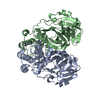
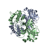
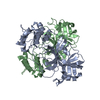
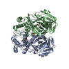
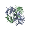
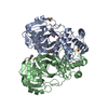
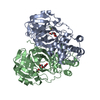
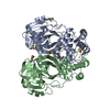
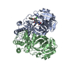
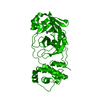
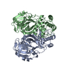
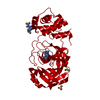
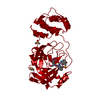
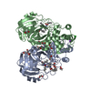
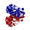
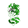
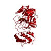
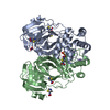
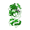
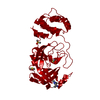
Entry Database : PDB / ID : 3f9hTitle Crystal Structure of the F140A mutant of SARS-Coronovirus 3C-like Protease at pH 7.6 3C-like proteinase Keywords / / / / / / / / / / Function / homology Function Domain/homology Component
/ / / / / / / / / / / / / / / / / / / / / / / / / / / / / / / / / / / / / / / / / / / / / / / / / / / / / / / / / / / / / / / / / / / / / / / / / / / / / / / / / / / / / / / / / / / / / / / / / / / / / / / / / / / / / / / / / / / / / / / / / / / / / / / / / / / / / / / / / / / / / / / / / / / / / / / / / / / / / Biological species Method / Resolution : 2.9 Å Authors Hu, T. / Li, L. / Jiang, H. / Shen, X. Journal : Virology / Year : 2009Title : Two adjacent mutations on the dimer interface of SARS coronavirus 3C-like protease cause different conformational changes in crystal structure.Authors : Hu, T. / Zhang, Y. / Li, L. / Wang, K. / Chen, S. / Chen, J. / Ding, J. / Jiang, H. / Shen, X. History Deposition Nov 13, 2008 Deposition site / Processing site Revision 1.0 Sep 29, 2009 Provider / Type Revision 1.1 Jul 13, 2011 Group Revision 1.2 Nov 10, 2021 Group / Category / struct_ref_seq_difItem / _database_2.pdbx_database_accession / _struct_ref_seq_dif.detailsRevision 1.3 Dec 27, 2023 Group / Category / chem_comp_bond
Show all Show less
 Movie
Movie Controller

 Yorodumi
 Open data
 Basic information
 Components
 Keywords
 Function and homology information
 SARS coronavirus
 X-RAY DIFFRACTION / Resolution: 2.9 Å
  Authors
 Citation
 Structure visualization
 Downloads & links
 Other downloads
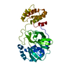


 PDBj
PDBj


 Assembly
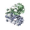

 Mass: 18.015 Da / Num. of mol.: 21 / Source method: isolated from a natural source / Formula: H2O
 Sample preparation
 Processing