Resolution: 2→29.501 Å / Num. obs: 28101 / % possible obs: 99.4 % / Observed criterion σ(I): -3 / Biso Wilson estimate: 23.871 Å2 / Rmerge(I) obs: 0.169 / Net I/σ(I): 7.58
Reflection shell
Resolution (Å)
Rmerge(I) obs
Mean I/σ(I) obs
Num. measured obs
Num. unique obs
Diffraction-ID
% possible all
2-2.07
0.01
1.9
22881
4849
1
95.9
2.07-2.15
0.01
2.6
28242
5007
1
99.8
2.15-2.25
0.01
3.2
30782
5326
1
99.8
2.25-2.37
0.01
4.1
30220
5225
1
99.8
2.37-2.52
0.01
5
30169
5203
1
99.8
2.52-2.71
0.01
6.6
29465
5066
1
99.9
2.71-2.99
0.01
8.3
30833
5324
1
100
2.99-3.42
0.01
11.6
29562
5112
1
99.9
3.42-4.3
0.01
15.3
29488
5150
1
99.7
4.3-29.501
0.01
17
29686
5202
1
99.4
-
Phasing
Phasing
Method: MAD
-
Processing
Software
Name
Version
Classification
NB
REFMAC
5.4.0067
refinement
PHENIX
refinement
SHELX
phasing
MolProbity
3beta29
modelbuilding
XSCALE
datascaling
PDB_EXTRACT
3.006
dataextraction
XDS
datareduction
SHELXD
phasing
autoSHARP
phasing
Refinement
Method to determine structure: MAD / Resolution: 2→29.501 Å / Cor.coef. Fo:Fc: 0.962 / Cor.coef. Fo:Fc free: 0.938 / Occupancy max: 1 / Occupancy min: 0.37 / SU B: 5.825 / SU ML: 0.086 / TLS residual ADP flag: LIKELY RESIDUAL / Cross valid method: THROUGHOUT / σ(F): 0 / ESU R: 0.132 / ESU R Free: 0.133 Stereochemistry target values: MAXIMUM LIKELIHOOD WITH PHASES Details: 1. HYDROGENS HAVE BEEN ADDED IN THE RIDING POSITIONS. 2. ATOM RECORDS CONTAIN RESIDUAL B FACTORS ONLY. 3. A MET-INHIBITION PROTOCOL WAS USED FOR SELENOMETHIONINE INCORPORATION DURING PROTEIN ...Details: 1. HYDROGENS HAVE BEEN ADDED IN THE RIDING POSITIONS. 2. ATOM RECORDS CONTAIN RESIDUAL B FACTORS ONLY. 3. A MET-INHIBITION PROTOCOL WAS USED FOR SELENOMETHIONINE INCORPORATION DURING PROTEIN EXPRESSION. THE OCCUPANCY OF THE SE ATOMS IN THE MSE RESIDUES WAS REDUCED TO 0.75 FOR THE REDUCED SCATTERING POWER DUE TO PARTIAL S-MET INCORPORATION. 4. CA ION, 2-METHYLPENTANE-2,4-DIOL AND ACETATE IONS HAVE BEEN MODELED FROM THE CRYSTALLIZATION CONDITIONS.
Rfactor
Num. reflection
% reflection
Selection details
Rfree
0.213
1408
5 %
RANDOM
Rwork
0.167
-
-
-
obs
0.169
28016
99.77 %
-
Solvent computation
Ion probe radii: 0.8 Å / Shrinkage radii: 0.8 Å / VDW probe radii: 1.2 Å / Solvent model: BABINET MODEL WITH MASK
In the structure databanks used in Yorodumi, some data are registered as the other names, "COVID-19 virus" and "2019-nCoV". Here are the details of the virus and the list of structure data.
Jan 31, 2019. EMDB accession codes are about to change! (news from PDBe EMDB page)
EMDB accession codes are about to change! (news from PDBe EMDB page)
The allocation of 4 digits for EMDB accession codes will soon come to an end. Whilst these codes will remain in use, new EMDB accession codes will include an additional digit and will expand incrementally as the available range of codes is exhausted. The current 4-digit format prefixed with “EMD-” (i.e. EMD-XXXX) will advance to a 5-digit format (i.e. EMD-XXXXX), and so on. It is currently estimated that the 4-digit codes will be depleted around Spring 2019, at which point the 5-digit format will come into force.
The EM Navigator/Yorodumi systems omit the EMD- prefix.
Related info.:Q: What is EMD? / ID/Accession-code notation in Yorodumi/EM Navigator
Yorodumi is a browser for structure data from EMDB, PDB, SASBDB, etc.
This page is also the successor to EM Navigator detail page, and also detail information page/front-end page for Omokage search.
The word "yorodu" (or yorozu) is an old Japanese word meaning "ten thousand". "mi" (miru) is to see.
Related info.:EMDB / PDB / SASBDB / Comparison of 3 databanks / Yorodumi Search / Aug 31, 2016. New EM Navigator & Yorodumi / Yorodumi Papers / Jmol/JSmol / Function and homology information / Changes in new EM Navigator and Yorodumi
 Movie
Movie Controller

 Yorodumi
 Open data
 Basic information
 Components
 Keywords
 Function and homology information
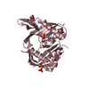
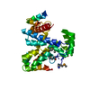
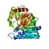
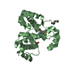
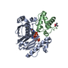
 Silicibacter sp. TM1040 (bacteria)
 X-RAY DIFFRACTION /
  Authors
 Citation
 Structure visualization
 Downloads & links
 Other downloads


 PDBj
PDBj
 Assembly


 Mass: 40.078 Da / Num. of mol.: 1 / Source method: obtained synthetically / Formula: Ca
 Mass: 118.174 Da / Num. of mol.: 1 / Source method: obtained synthetically / Formula: C6H14O2 / Comment: precipitant*YM
 Mass: 118.174 Da / Num. of mol.: 6 / Source method: obtained synthetically / Formula: C6H14O2 / Comment: precipitant*YM
 Mass: 59.044 Da / Num. of mol.: 2 / Source method: obtained synthetically / Formula: C2H3O2
 Sample preparation
 / Beamline: 23-ID-B / Wavelength: 0.97967,0.94645
 Processing